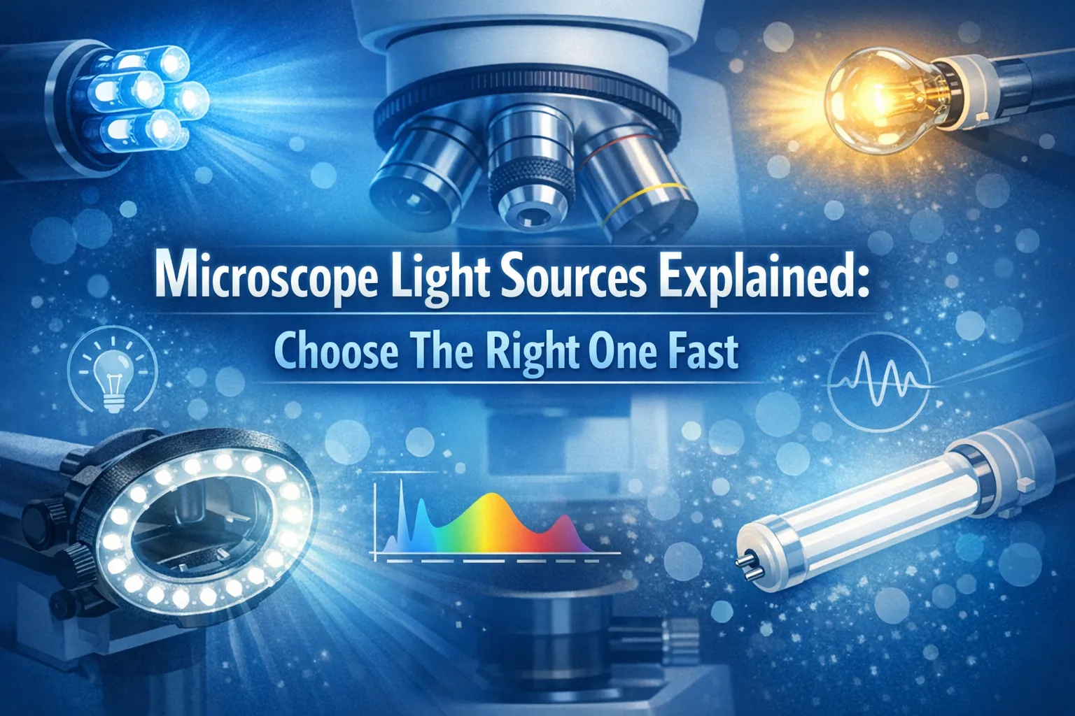
You need the right microscope light source for clear results. LED, halogen, fluorescent, fiber optic, arc-lamp, and UV each fit different tasks. LED provides stable brightness and suits long experiments. Halogen works for routine imaging. Fluorescent and arc-lamp excel in fluorescence applications. Fiber optic delivers intense light for stereo microscopes. UV and lasers support specialized studies. Matching your light source to your application improves imaging quality, protects samples, and saves costs. Instant on/off switching reduces phototoxicity when you observe live cells.
LEDs last longer and use less energy than traditional lamps. Heat and vibration from halogen or arc-lamp can affect sensitive samples.
Choose your light source based on your imaging needs, intensity control, and safety.
Next, compare each option to find the best fit for your workflow.
Key Takeaways
-
Match your light source to your application for better imaging quality, cost savings, and sample protection. LEDs are ideal for long experiments, while arc-lamps excel in fluorescence microscopy.
-
LEDs offer the longest lifetime, energy efficiency, and stable brightness, making them a top choice for routine and advanced imaging tasks.
-
Consider safety and compatibility when choosing a light source. LEDs generate minimal heat, while arc-lamps require careful handling due to high intensity and heat production.
Microscope Light Source Comparison
LED, Halogen, Fluorescent, Fiber Optic, Arc-Lamp, UV
You need to compare each microscope light source to make the best choice for your microscopy workflow. The main types include LED, halogen, fluorescent, fiber optic, arc-lamps, and UV. Each light source offers unique illumination characteristics, spectrum coverage, and intensity control.
You can see the differences in the table below, which summarizes light output and spectral range for each option:
|
Light Source |
Light Output Characteristics |
Spectral Range Characteristics |
|---|---|---|
|
LEDs |
Energy-efficient, long-lasting, low-heat, stable illumination |
Emission peak at 460 nm (blue), broad range 550-650 nm |
|
Halogen lamps |
Bright, broad-spectrum light with good color rendering |
Good color rendering across visible spectrum |
|
Metal halide lamps |
High-intensity, broad-spectrum light |
Suitable for fluorescence imaging |
|
Mercury vapor lamps |
Intense light at specific wavelengths |
Peaks in 300-450 nm range |
|
Xenon arc-lamps |
Broader intensity output across visible spectrum |
Deficient in UV, high intensity in infrared |
|
Arc-lamps |
10-100 times brighter than tungsten lamps |
Intensity peaks in near-UV and blue spectrum |
You can use this table to quickly identify which light source fits your application. For example, widefield fluorescence microscopy often requires high-intensity light and specific excitation wavelengths, so arc-lamps and metal halide lamps are common choices. LED and halogen lamps work well for routine imaging and photomicrography, while fiber optic sources deliver intense illumination for stereo microscopes.
Pros, Cons, and Best Use Cases
You need to weigh the advantages and disadvantages of each light source before making a decision. Here is a quick overview:
|
Light Source |
Advantages |
Disadvantages |
|---|---|---|
|
LEDs |
Longest lifetime, stable brightness, energy-efficient, no warm-up time, precise intensity control, safer (no UV or toxic chemicals) |
High upfront cost, weaker intensity in green excitation region, limited UV excitation capability. |
|
Arc-lamps |
Inexpensive to buy, suitable for UV excitation, consistent intensity (xenon arc-lamps) |
Short lifetime, hazardous waste disposal, uneven illumination, high heat production, photobleaching risk. |
|
Tungsten-halogen |
Relatively cheap, pre-aligned, broad wavelength range, suitable for visible light experiments. |
Shorter lifespan than LEDs, emits excess heat, weak UV emission, requires heat/UV filters. |
-
LED illumination microscopes are preferred for their efficiency and performance. You get bright, even light that enhances clarity and detail. Many users highlight the importance of stable illumination for optimal viewing of specimens. Proper lighting can improve visibility by over 40%, making LED options a popular choice.
-
Halogen lights provide great color integrity, which is important for photomicrography and research. You can easily replace bulbs, but you must handle them carefully to avoid damage. Halogen bulbs produce white light when new but shift to yellow as they age.
-
Arc-lamps deliver high intensity and are ideal for widefield fluorescence microscopy. You must consider safety precautions because these lamps generate extreme heat and require careful handling.
Performance, Cost, Lifetime, Safety
You need to understand how each light source performs in terms of energy efficiency, heat generation, cost, lifetime, and safety.
Tip: LED lights achieve 80%-90% efficiency, generating minimal heat and maximizing light output. Halogen lights convert only 10%-20% of energy into light, wasting the rest as heat. This increases electricity consumption and cooling costs.
-
LED lights last up to 50,000 hours, reducing long-term costs and minimizing downtime. Halogen bulbs last up to 2,000 hours, while fluorescent light sources reach up to 10,000 hours. You can expect LED to offer the longest lifetime and lowest maintenance.
-
Arc-lamps, including mercury and xenon arc-lamps, require caution. You must avoid direct observation of the burning lamp to prevent eye damage. Always allow sufficient cooling time before changing bulbs. Store lamps in their shipping containers to prevent accidents. Never touch lamps with bare fingers, as this can etch the quartz envelope.
-
LED lights do not heat up as much as halogen bulbs, making them safer for sensitive specimens. However, LED lights may cause color distortion in some samples, which can affect photomicrography results.
-
Arc-lamps produce high-intensity light and are suitable for excitation in fluorescence applications. You must manage photobleaching risk and uneven illumination. These lamps have short lifetimes and require hazardous waste disposal.
You can select the best microscope light source by considering your application, required intensity, spectrum coverage, and safety needs. For widefield fluorescence microscopy, you need high-intensity light and precise excitation wavelengths. LED and halogen lamps work well for routine imaging and photomicrography.
Fiber optic sources provide intense illumination for stereo microscopes. You must match the light source to your workflow to achieve optimal results in microscopy.
Match Light Source to Application
Choosing the right microscope light source for your application ensures you achieve optimal results in microscopy. You must consider illumination, intensity, spectrum, compatibility, and safety. Each imaging technique demands specific features from its light source. You can match your selection to fluorescence, brightfield, stereo, or live cell imaging by understanding what each method requires.
Fluorescence and Fluorescent Light Sources
Fluorescence microscopy relies on powerful illumination and precise excitation wavelengths. You need a microscope light source that delivers high-intensity light to excite fluorophores and generate detectable emissions. The most suitable options for fluorescence include:
-
LEDs: You benefit from long lifetime, stable brightness, and consistent excitation conditions. LEDs work well for automated experiments and widefield fluorescence microscopy. They require no warm-up time and offer low maintenance.
-
Tungsten-halogen lamps: These lamps provide a broad spectrum of visible wavelengths. You can use them for experiments needing a wide range of light, but they emit excess heat and have weak UV emission. They are less suitable for imaging below 400nm.
-
Arc-lamps: Arc-lamps, including mercury and xenon arc-lamps, deliver strong UV emission and high-intensity light. You use them for applications requiring excitation below 400nm. They are less common today due to shorter lifetime and higher maintenance.
Although LEDs have a higher upfront cost, you save money over time because of their low maintenance, energy efficiency, and stable illumination. You do not need to align or dispose of LEDs as hazardous waste. LEDs are more economical for widefield fluorescence microscopy and photomicrography.
In fluorescence, the light source must emit specific wavelengths for excitation. You need high intensity to maximize emission.
White light sources contain all visible spectrum wavelengths, allowing you to select excitation filters for precise excitation. However, you must ensure high intensity in the excitation spectra to prevent unwanted excitation and light damage.
|
Light Source Type |
Wattage Range |
Key Characteristics |
|---|---|---|
|
Mercury Burner |
50 – 200 watts |
Peaks at specific wavelengths, high intensity in near UV |
|
Xenon Burner |
75 – 150 watts |
Provides broad spectrum, powerful light source for detection |
You must handle arc-lamps carefully. Mercury and xenon arc-lamps operate under high pressure and require safety precautions. You avoid direct exposure to the burning lamp and allow cooling before changing bulbs. You must store lamps properly and never touch them with bare fingers.
Brightfield, Stereo, and Routine Imaging
Brightfield and stereo microscopy require stable illumination and accurate color rendering. You select a microscope light source based on spectrum coverage and intensity. LEDs and halogen lamps are the most common choices for these applications.
-
LEDs: You gain energy efficiency, long lifetime, and stable illumination. LEDs work well for both stereo and fluorescence microscopy. You avoid excess heat, which protects specimens and microscope components.
-
Halogen lamps: You receive bright, broad-spectrum light with excellent color rendering. Halogen lamps are ideal for brightfield and stereo microscopy. You can easily replace bulbs and maintain consistent illumination.
-
For stereo microscopes: You use LED ring lights, gooseneck illuminators, and fiber optic light guides. These options enhance visibility of three-dimensional specimens and provide high-intensity light.
Color rendering impacts image clarity in routine microscopy and photomicrography. You must choose a light source with a high CRI (Color Rendering Index) value to ensure true color images.
|
Description |
Impact on Microscopy |
|
|---|---|---|
|
100 |
True colors as under natural sunlight |
Ideal for color-sensitive applications |
|
90+ |
High accuracy in color representation |
Ensures full wavelength spectrum for true color images |
|
Low CRI |
Weak in deep red and cyan/blue-green |
Colors appear washed out and distorted, affecting clarity and contrast |
You achieve the best results in photomicrography by selecting a light source with high CRI and stable illumination. You avoid color distortion and maintain clarity in your images.
Compatibility matters when you choose a microscope light source. Arc-lamps and tungsten-halogen lamps generate heat and vibration, which can damage samples and disrupt imaging quality. You use liquid light-guides to deliver high-intensity light from a remote source, reducing electrical noise and vibration at the microscope.
Although coupling losses may reduce light power, the high intensity compensates for this and ensures effective performance.
Live Cell Imaging and Intensity Control
Live cell imaging requires careful management of illumination and intensity. You must protect live cells from photobleaching and maintain cell viability. You avoid high light intensities and long exposure times to ensure specimen health.
-
You optimize spatial and temporal resolution to match your experimental goals. You prevent oversampling and overexposing cells.
-
You use motorized shutters, filter wheels, and focus control mechanisms to enhance efficiency and precision. These features support time-lapse experiments and minimize light exposure.
-
You select a microscope light source with precise intensity control and stable illumination. LEDs and light-emitting diodes are preferred for their low heat generation and consistent brightness.
Tip: Maintaining cell viability is crucial during live-cell imaging experiments. You avoid high-intensity light and long exposures to prevent photobleaching and ensure specimen health.
Safety certifications are essential for microscope light sources. You look for IEC 62471 certification for photobiological safety and ANSI Z136 standards for laser safety. These certifications ensure safe exposure levels and protect you during microscopy. You match your microscope light source to your application by considering illumination, intensity, spectrum, compatibility, and safety.
You achieve optimal results in widefield fluorescence microscopy, photomicrography, and live cell imaging by selecting the right light source for your workflow.
Fast Light Source Selection Tips
Quick Checklist for Choosing
You can streamline your decision-making process by following a practical checklist. This approach helps you select the best light source for your microscopy needs. Consider these key factors:
-
Identify your application: Are you working with widefield fluorescence microscopy, photomicrography, or routine imaging?
-
Determine the required illumination and intensity for your specimens.
-
Check the compatibility of the light source with your microscope. Direct mounting reduces heat and vibration, while remote sources with liquid light-guides minimize interference.
-
Evaluate the spectrum coverage and wavelengths needed for excitation and emission. Use excitation filters to target specific wavelengths.
-
Compare the average lifetime and maintenance requirements. LEDs offer stable brightness and long lifetime. Arc-lamps provide high intensity but require frequent replacement.
-
Assess safety features, especially for arc-lamps and xenon arc-lamps. Look for certifications and proper handling instructions.
A structured checklist ensures you consider all relevant options. You avoid missing important details and make informed choices for optimal illumination and intensity.
Common Mistakes to Avoid
You can prevent common pitfalls by staying alert to these issues:
-
Forgetting to set the rheostat to the highest brightness level.
-
Leaving the field iris closed, which blocks illumination.
-
Engaging the beam splitter on a trinocular microscope, reducing light intensity.
-
Misplacing the disc diaphragm, affecting spectrum and illumination.
-
Ignoring bulb burnout or fuse damage, which shortens average lifetime.
-
Using uncharged batteries in cordless models.
-
Overlooking heat and vibration from arc-lamps, which can damage microscope parts and affect fluorescence imaging.
You can choose the right light source quickly by matching it to your application. For fluorescence, arc-lamps offer high intensity at specific wavelengths. Arc-lamps also suit advanced imaging, but you must consider safety. Use the comparison and checklist to weigh arc-lamps against other options and select confidently.
FAQ
What is the safest light source for sensitive specimens?
You can use LED lights. They produce minimal heat and stable illumination. This protects your samples and reduces risk during microscopy.
What light source works best for fluorescence microscopy?
You should select arc-lamps or LEDs. These provide high-intensity light and specific wavelengths needed for fluorescence imaging.
What is the average lifetime of microscope light sources?
LEDs last up to 50,000 hours. Halogen bulbs last about 2,000 hours. Arc-lamps usually last between 300 and 2,000 hours.

