When you face an imaging or research challenge, choosing the right microscope becomes crucial. You should focus on how much resolution and magnification your work demands. If you need to observe fine details, like the ultrastructural morphology of cells or tiny particles, you might benefit from advanced techniques such as correlative light and electron microscopy or three-dimensional imaging.
For tasks like diagnosing glomerular diseases or art conservation—where precise resolution and magnification are essential—electron microscopes often provide the clarity you need.
Consider your budget, the type of sample you plan to examine, and whether you want to view live processes in real time. The electron microscope vs light microscope decision often comes down to these factors:
-
The level of resolution required for your research
-
The magnification needed to see your sample clearly
-
The complexity of sample preparation
-
The importance of observing live or dynamic samples
With these points in mind, you can move forward to see how light microscopes offer practical solutions for many everyday imaging needs.
Light Microscope Solutions
Everyday Imaging Needs
You can use a light microscope for a wide range of everyday imaging tasks. This tool uses visible light to illuminate your samples, making it easy to observe cells, tissues, and even small organisms. You do not need advanced training to operate a light microscope. You can quickly set up your sample and start viewing it within minutes. The light microscope allows you to see colored images, which helps you distinguish between different cell types and structures.
If you want to study sub-cellular structures, this microscope provides enough detail for most classroom and laboratory needs.
Cost and Accessibility
A light microscope offers an affordable solution for most educational and research settings. You can find basic models for a few hundred dollars, and even advanced versions rarely exceed a few thousand dollars. In contrast, electron microscopes can cost from $30,000 for tabletop models to several million dollars for high-end systems.
The table below compares the price ranges:
|
Type of Microscope |
Price Range |
|---|---|
|
Light Microscope |
$100 to $10,000 |
|
Scanning Electron Microscope (SEM) |
$80,000 to $2,000,000 |
|
Transmission Electron Microscope (TEM) |
$300,000 to $10,000,000 |
|
Focused Ion Beam Electron Microscope (FIB) |
$500,000 to $4,000,000 |
You can also maintain a light microscope at a low cost. This makes it accessible for schools, clinics, and small labs.
Live Sample Observation
You can observe living organisms with a light microscope. This feature sets it apart from electron microscopes, which require complex preparation that kills the sample. You can watch cells move, divide, and interact in real time. This ability supports live-cell imaging and helps you study biological processes as they happen. The light microscope also allows you to view thicker samples and produces color images, making it ideal for observing dynamic changes in living specimens.
Electron Microscope Solutions
High-Resolution Imaging
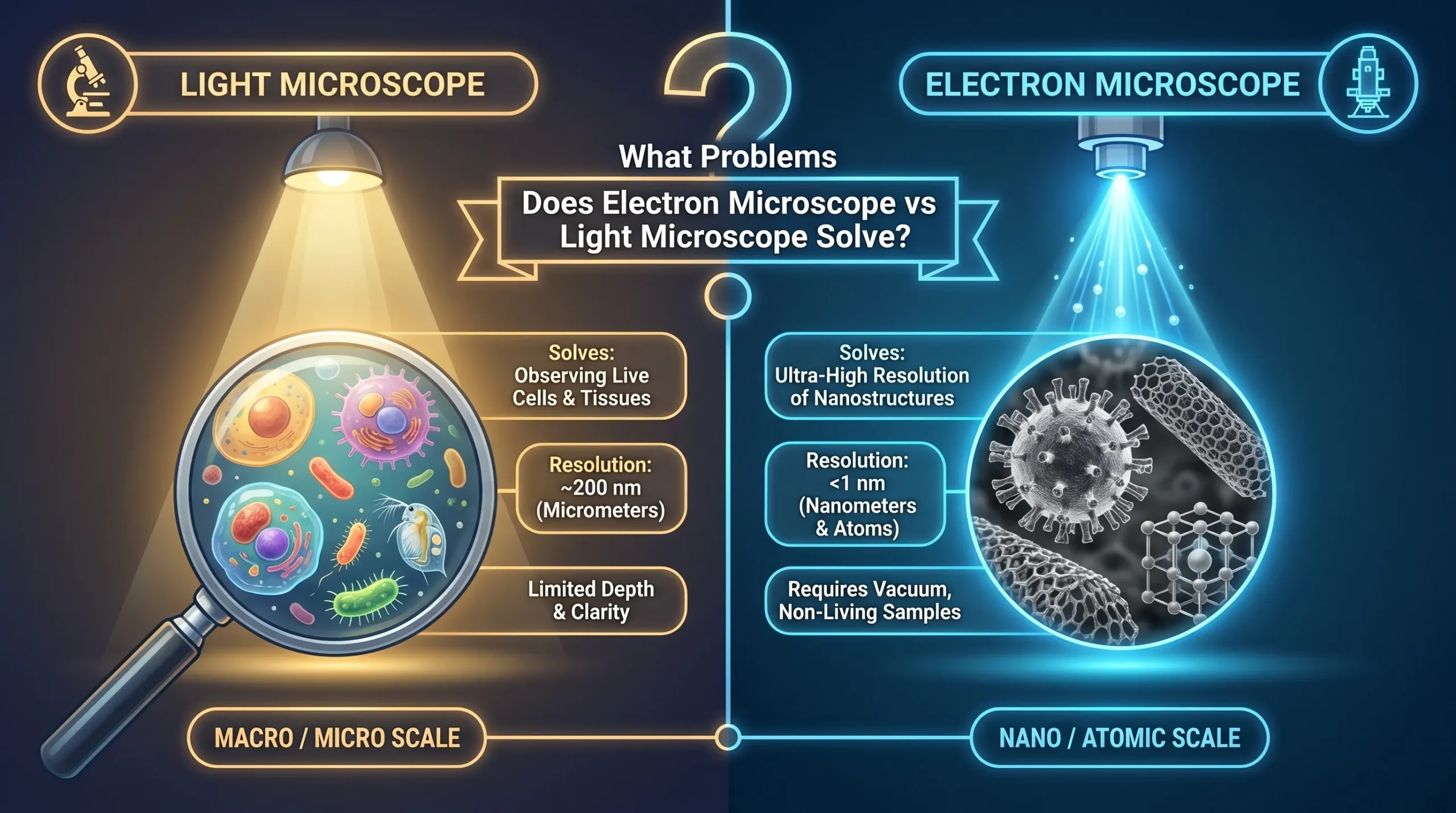
You can use an electron microscope when you need high-resolution imaging that surpasses the limits of light microscopes. Electron microscopes use beams of electrons instead of visible light, which gives you much higher magnification and resolution. Modern electron microscopes achieve a resolution of about 0.20 nm, while light microscopes are limited to around 200 nm. This means you can see details that are 250 times smaller than what a light microscope can reveal.
The high-resolution performance of electron microscopy allows you to observe the ultrastructure of cells, viruses, and even individual molecules. If you need sub-nanometer imaging, electron microscopes provide the capabilities you require for detailed imaging at the atomic level.
|
Microscope Type |
Resolution |
|
|---|---|---|
|
Light Microscope |
Up to 1,000x |
~200 nm |
|
Electron Microscope |
Up to 1,000,000x |
0.1 nm |
Structural Detail Analysis
Electron microscopy gives you access to high-resolution analysis of structures that light microscopes cannot resolve. You can visualize the honeycomb lattice of graphene, the folds of viral capsids, and the double helix of DNA. These details are essential for ultrastructure analysis in biology, materials science, and nanotechnology. The high-resolution capabilities of electron microscopes let you study the ultrastructure of cells, including organelles and membranes, with unmatched clarity.
You can use electron microscopy to perform ultrastructure analysis in cellular biology, microbiology, and materials research.
Small Particle Visualization
You can use an electron microscope to visualize particles much smaller than those seen with a light microscope. Electron microscopes allow you to see viruses, proteins, and even glucose molecules. The diffraction limit of visible light restricts light microscopes to structures larger than 200 nm, but electron microscopes overcome these resolution limits. With high-performance benchtop SEMs and advanced TEMs, you can achieve magnification up to 1,000,000x and observe ultrastructure at the nanometer and sub-nanometer scale.
Electron microscopy enables detailed imaging of the smallest particles, supporting research in virology, nanotechnology, and materials science.
-
Electron microscopes visualize structures smaller than 1 nm.
-
You can analyze viruses (30-250 nm), proteins (10 nm), and molecules (1 nm).
-
Light microscopes cannot resolve these ultrastructure details.
The electron microscope vs light microscope comparison shows that electron microscopes solve the problem of high-resolution imaging, ultrastructure analysis, and small particle visualization. You gain access to advanced capabilities and performance for detailed imaging, though you must prepare your specimens carefully.
Electron Microscope vs Light Microscope: Choosing the Right Tool
Match Microscope to Your Needs
When you decide between an electron microscope vs light microscope, you need to match the tool to your research goals. Each microscope solves different problems based on what you want to see and how you want to observe it. If you want to study living cells or tissues in real time, a light microscope gives you the flexibility to watch biological processes as they happen. You can use it for quick checks, classroom demonstrations, or routine lab work.
The light microscope works well for microbiology and pathology, where you need to see cells, tissues, or microorganisms with color and minimal preparation.
If your research requires you to see structures at the molecular or atomic level, you need the power of an electron microscope. This tool lets you explore the ultrastructure of cells, viruses, and nanomaterials. You can achieve much higher magnification and resolution, which is essential for fields like virology, molecular biology, neuroscience, and materials science. The electron microscope helps you solve problems that need detailed imaging of tiny particles or complex structures.
You should also consider your budget, the type of sample, and how much time you have for preparation. The electron microscope costs more to buy and maintain, and it requires a controlled environment. The light microscope is more affordable and easier to use in most settings.
Recent advances have made electron microscopes easier to operate, but you still need to prepare your samples carefully, and you can only use dead specimens.
The table below summarizes the main factors to help you choose the right microscope for your needs:
|
Factor |
Light Microscope |
Electron Microscope |
|---|---|---|
|
Research Focus |
Best for observing live cells and basic structures like tissues and microorganisms. |
Necessary for high-resolution imaging of cellular ultrastructure and nanomaterials. |
|
Sample Preparation |
Quick and easy, suitable for live or minimally processed samples. |
Complex and time-consuming, requiring fixing and coating with heavy metals. |
|
Budget |
Generally affordable with low operating costs. |
Expensive to purchase and maintain, requiring specialized environments. |
|
Imaging Needs |
Ideal for real-time observation and colored images. |
Provides high-resolution grayscale images, requiring interpretation. |
Decision Checklist
You can use a simple checklist to match your research needs to the right microscope. Ask yourself these questions before you choose:
-
What type of sample do you need to examine—cultured cells, tissue sections, or whole organisms?
-
Do you need to observe live specimens or only dead ones?
-
What level of magnification and resolution do you require for your study?
-
How much time can you spend on sample preparation?
-
Is your budget sufficient for high-end equipment and maintenance?
-
Do you need real-time observation or detailed structural analysis?
-
Which research field are you working in—microbiology, pathology, virology, materials science, or neuroscience?
You can also compare the key features of each microscope type in the table below:
|
Feature |
Light Microscope |
Electron Microscope |
|---|---|---|
|
Magnification |
Up to 1,500x |
Up to 1,000,000x |
|
Resolution |
~200 nm |
~0.1 nm |
|
Specimen Preparation |
Simple and quick |
Labor-intensive, requires extensive preparation |
|
Observation |
Direct viewing, real-time |
Projected images, grayscale |
|
Cost |
Low |
High |
|
Maintenance |
Low |
High |
|
Sample Type |
Living/Dead |
Dead Only |
|
Accessibility |
High |
Limited |
You should also think about the time needed for sample preparation. Light microscopes let you prepare samples in minutes or hours, while electron microscopes often require several days. The environment also matters. Light microscopes work at atmospheric pressure and support live-cell imaging. Electron microscopes need a vacuum and sometimes special coatings for samples.
Here is a summary of the main differences:
-
Magnification and resolution: Light microscopes reach up to 1,500x and about 200 nm, while electron microscopes go up to 1,000,000x and about 0.1 nm.
-
Sample preparation: Light microscopes need simple, quick preparation. Electron microscopes require more steps and only work with dead samples.
-
Observation: Light microscopes give you real-time, color images. Electron microscopes provide grayscale images that show more detail but need interpretation.
-
Cost and accessibility: Light microscopes are affordable and easy to use. Electron microscopes are expensive and need special environments.
By using this checklist and comparing the features, you can confidently choose the right microscope for your research or educational needs.
When you compare electron microscope vs light microscope, you solve different problems based on your needs. Light microscopes help you with quick, affordable imaging and live sample observation. Electron microscopes give you unmatched resolution for detailed research. Consider what you want to see, your budget, and how much detail you need.
-
Use a light microscope for general observations and education.
-
Choose an electron microscope for advanced analysis and high-resolution imaging.
FAQ
What can you see with a light microscope?
You can see cells, tissues, bacteria, and some small organisms. You also view colored images and observe living samples in real time.
What makes an electron microscope different from a light microscope?
You use an electron microscope to see much smaller details. It uses electron beams, not light, and gives you higher magnification and resolution.
What samples work best for electron microscopy?
You should use very thin, dry, and non-living samples. Electron microscopes work best for viruses, cell structures, and tiny particles.
What are the main limitations of a light microscope?
You cannot see structures smaller than 200 nanometers. You also get less detail and lower magnification compared to electron microscopes.
What should you consider before choosing a microscope?
You need to think about your sample type, the level of detail you want, your budget, and if you need to observe live samples.

