Imagine a secret universe, invisible to the naked eye—where tiny creatures swim, crawl, and multiply right under your nose. With a microscope, you become an explorer of this hidden realm. Peek through the lens and discover bustling colonies of bacteria or delicate forests of fungi. It does not matter if you’re using prepared slides or creating your own from everyday items, the adventure begins with a single drop.
Grab a microscope, place your sample on the stage, and uncover the amazing world that science reveals just beneath the surface
What Are Germs?
You hear about germs a lot. But what are germs? Germs are tiny living things. They can make you sick. You cannot see germs without a microscope. If you put something under a microscope, you see a hidden world. There are strange shapes and colors. Germs live everywhere. They are on your hands. They float in the air. They can be on your favorite snack.
You need a microscope to see these micro monsters. They are much smaller than what your eyes can see.
Types of Germs: Bacteria, Viruses, Fungi
Let’s look at the main types of germs. You might find these when you use a microscope. Each type is different. They have their own shape and size. They live in their own way.
Here is a table to help you compare them:
|
Category |
Definition |
Characteristics |
Size Range (micrometers) |
Example Diseases |
|---|---|---|---|---|
|
Bacteria |
Unicellular prokaryotic microorganisms that lack a true nucleus and membrane-bound organelles. |
Unicellular, prokaryotic, have cell walls, reproduce by splitting, live almost everywhere. |
0.2–2.0 |
Strep throat, pneumonia |
|
Viruses |
Extremely small infectious particles that depend on a host cell for survival and reproduction. |
Not alive outside a host, contain DNA or RNA, infect all life forms, need a microscope with high power. |
0.02–0.3 |
Flu, COVID-19 |
|
Fungi |
Eukaryotic organisms, can be unicellular (yeast) or multicellular (mold). |
Have cell walls, grow as filaments or single cells, can be seen with a basic microscope. |
2–10 |
Athlete’s foot, ringworm |
Did you know? Scientists think one gram of soil can have up to 40 million bacterial cells (source). That is a lot of things to look at with your microscope!
When you use a microscope, you might see bacteria. They look like tiny rods or balls. Viruses are much smaller. You need a very strong microscope to see them. Fungi can look fuzzy or like small ovals. Each thing you look at shows you something new in the micro world.
Why Germs Are Invisible to the Naked Eye
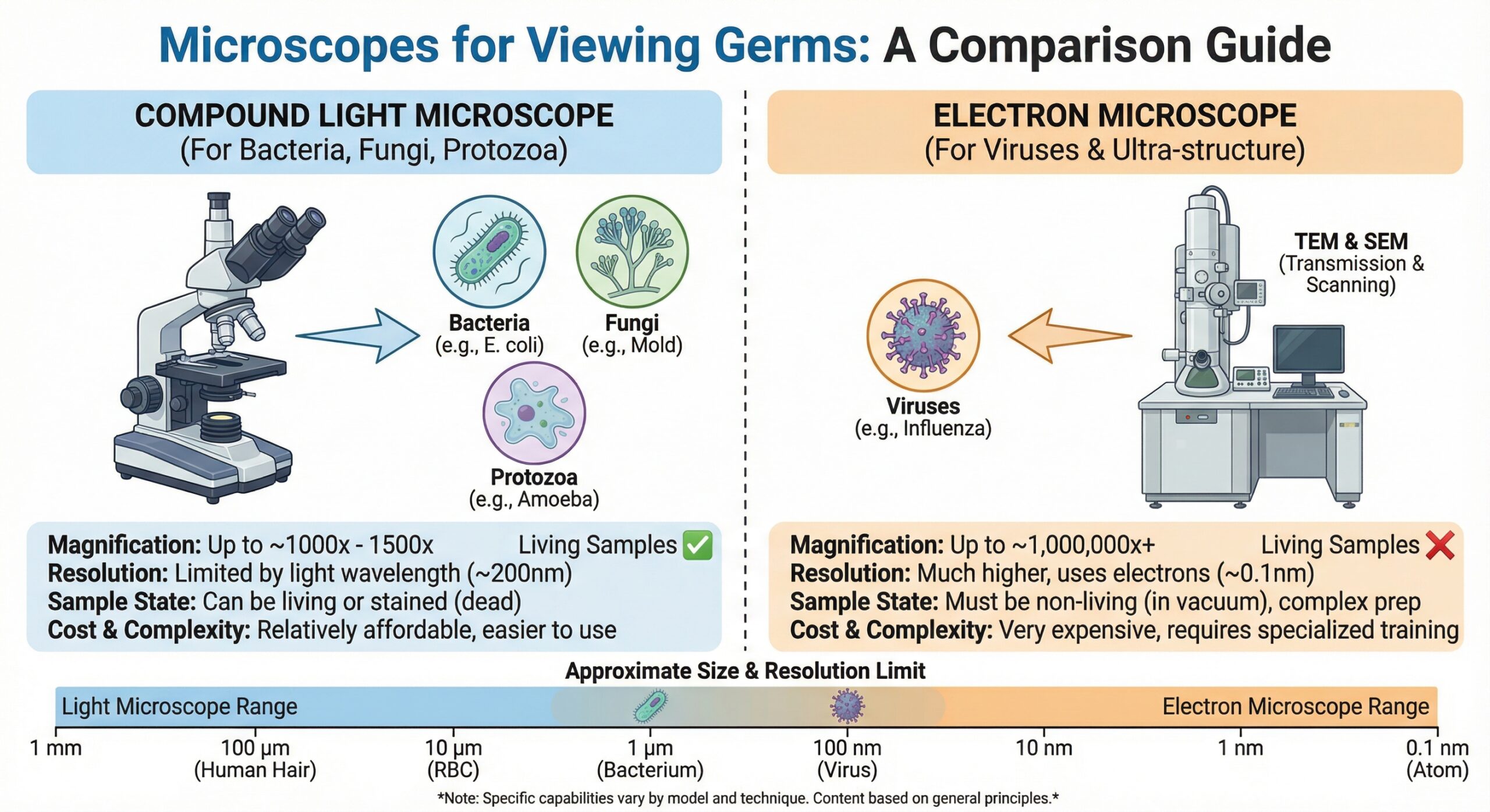
You cannot see germs with your eyes. Germs are much smaller than anything you can spot. Your eyes need light to bounce off things. Germs are so tiny that light does not bounce enough for you to see them. Most bacteria are less than two micrometers wide. Viruses are even smaller. Some are only 0.02 micrometers wide. Fungi are bigger, but you still need a microscope to see them well. If you put a specimen on a slide and look through a microscope, you finally see what has been hiding.
If you want to see germs, you need a microscope with good magnification. Most classroom microscopes can show you bacteria and fungi. You need a special electron microscope to see viruses. Each thing you look at helps you learn why washing your hands and keeping things clean is important.
How a Microscope for Germs Works
Magnification and Lenses Needed to See Germs
To see germs, you need a microscope with enough power. Not every microscope can show all germs. You might ask, “How strong should my microscope be?”
Let’s explain it.
Most classrooms use a compound microscope. This tool helps you look at bacteria and fungi. These microscopes can zoom from 40x to 1000x. With this, you can see bacteria as small rods or balls. Fungi look fuzzy or like ovals. Put your sample on a glass slide. Focus the lens. You will see the micro monsters appear. Viruses are much smaller than bacteria or fungi. You need an electron microscope to see them.
Electron microscopes use beams of electrons, not light. They can zoom over 1,000,000x! The first virus photo was taken in 1938 with this tool. New electron microscopes, like cryo-electron ones, can even show single atoms (source). That is how you see the tiniest germs.
Here is a table to help you compare what you need for each germ:
|
Germ Type |
Typical Size (micrometers) |
Recommended Microscope |
Magnification Needed |
Lens Type |
Special Techniques |
|---|---|---|---|---|---|
|
Bacteria |
0.2–2.0 |
400x–1000x |
Glass objective |
Gram stain, acid-fast |
|
|
Fungi |
2–10 |
Compound microscope |
100x–400x |
Glass objective |
Wet mount |
|
Viruses |
0.02–0.3 |
Electron microscope |
50,000x–1,000,000x |
Electron lens |
Negative staining |
|
Protozoa |
10–50 |
Compound microscope |
100x–400x |
Glass objective |
Simple stain |
You must prepare your sample the right way. Use stains like Gram or acid-fast to make bacteria easier to see. Put your sample on a slide. Add a drop of water. Cover it with a coverslip. Adjust the light and focus to see details. Light microscopes are good for bacteria and fungi. Electron microscopes let you see viruses and tiny cell parts. Microscope for Germs: Choosing the Right Tool
Picking a microscope for germs is like choosing a cool gadget. You want to see tiny germs up close. You also need a microscope that works for you.
Let’s look at the choices so you can pick the best one for your science fun.
Light Microscopes for Germs
You probably use light microscopes in your science class. These microscopes use glass lenses and light to make things look bigger. You can see bacteria and fungi with a compound microscope. To see bacteria better, use the gram stain method. This method adds color to bacteria so you can tell them apart. Put a drop of stain on your sample, wait, and rinse it off. Then, purple and pink bacteria show up under the lens!
Light microscopes are good for slides that are already prepared. Many science kits have slides with germs from things at home. Some microscopes for germs are made for kids. They have easy knobs and strong frames. You can even use a home microscope with slides from your kitchen or bathroom.
If you want to see tuberculosis bacteria, use the acid-fast stain. This special dye helps you find germs that regular stains miss.
Here’s a quick list of things to think about when picking a microscope for germs:
|
Feature |
Description |
|---|---|
|
Sample Type |
Use upright compound microscopes for prepared slides and inverted microscopes for live culture observation. |
|
Contrast Method |
Phase contrast is ideal for transparent microbes, while fluorescence is essential for tagged microbial studies. |
|
Optical Quality |
Infinity plan objectives ensure sharp, distortion-free imaging required in research applications. |
|
Digital Documentation |
Choose trinocular or digital-ready models for recording, analyzing, and sharing microbial research. |
|
Budget & Flexibility |
Basic phase contrast microscopes meet standard microbiology needs, while modular fluorescence systems provide advanced imaging capabilities. |
Electron Microscopes for Tiny Details
Sometimes you want to see germs even smaller than bacteria. Viruses are so tiny that you need an electron microscope to see them. These microscopes use electron beams instead of light. You can zoom in up to a million times! Transmission electron microscopy (TEM) lets you see the shape and size of viruses. For example, scientists use TEM to look at viruses like bovine herpesvirus, which are about 150 to 200 nanometers wide (source). That is much smaller than bacteria.
Electron microscopes help you see the smallest parts of your sample. You can see the shape of viruses, cell walls, and even inside bacteria. These microscopes for germs are very strong, but they cost a lot and are hard to use. Most homes and schools do not have them, but you can see their pictures in books or online.
Digital Microscopes for Easy Viewing
Digital microscopes make looking at germs easy and fun. You connect them to a computer or tablet and see your sample on a big screen. You can take pictures, record videos, and share what you find with friends or teachers. Many digital microscopes for germs are simple to use, so you do not need to be a scientist. Some even let you join online science labs from home.
Schools like digital microscopes because they are cheap and simple to use. You can zoom in, change the light, and take pictures for your project. New features like comfy designs and digital pictures make these microscopes great for school and home. You can look at germs from your kitchen, bathroom, or backyard with just a few clicks. Here is a table that compares light, electron, and digital microscopes for germs:
|
Type of Microscope |
Magnification Range |
Best For |
Accessibility |
Unique Features |
Citation |
|---|---|---|---|---|---|
|
Light |
40x–1000x |
Bacteria, fungi |
High (schools, home) |
Uses stains like gram and acid-fast |
|
|
Electron |
50,000x–1,000,000x |
Viruses, cell details |
Low (labs only) |
Reveals viral particles, cell ultrastructure |
|
|
Digital |
40x–1000x |
Bacteria, fungi, protozoa |
Very high (all ages) |
Connects to devices, supports remote learning |
Let’s see what makes a microscope for germs good for learning:
|
Factor |
Description |
|---|---|
|
Cost |
Schools and families want affordable microscopes for germs so everyone can explore. |
|
Accessibility |
User-friendly designs help kids and adults use microscopes without trouble. |
|
Technological Advancements |
Digital imaging and ergonomic shapes make microscopes more fun and easier to use. |
|
Remote Learning Support |
Features for online labs let you study germs from anywhere. |
You have lots of choices when picking a microscope for germs. You can use a compound microscope with gram stain for bacteria. You can use an electron microscope for viruses. You can use a digital microscope for easy sharing. Each tool lets you see something new in your sample.
Seeing Germs Under the Microscope
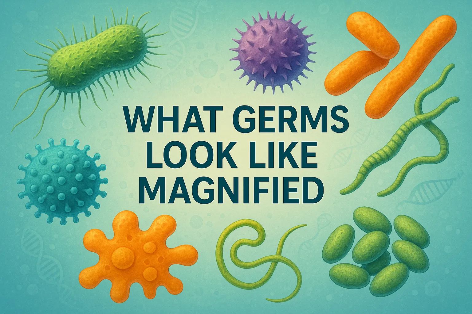
What Germs Look Like Magnified
Looking through a microscope feels like entering a new world. You see shapes and colors you never saw before. Bacteria can look like tiny rods, balls, or spirals. Some stick together, but others float by themselves. Fungi look like fuzzy threads or small ovals. A compound microscope helps you spot these microbe shapes. Science kits have slides that show bacteria from places like your kitchen or bathroom.
Sometimes, bacteria turn purple or pink after using the gram stain. This color change helps you tell them apart.
If you want to see more details, try using stains. The gram stain makes bacteria easier to see. The acid-fast stain helps you spot germs like tuberculosis. Take a sample from home and put it on a slide. Add a drop of water and cover it with a coverslip. Change the focus and light. Suddenly, you can see the micro monsters moving around.
Common Experiments: Soap, Hand Hygiene, and More
You can do easy experiments to see how germs move and how cleaning helps. These activities make science fun and show why hygiene is important. Get your microscope for germs, some slides, and things from home. Here are some experiments you can try:
-
Hand Hygiene Test
Rub your finger on a slide before washing your hands. Look at it under the microscope. Wash your hands with soap, then rub your finger on a new slide. Compare both slides. You will see fewer bacteria after washing. -
Soap Effectiveness
Touch a bar of soap, then press your finger on a slide. Use the microscope to look for microbes. Try the same thing with liquid soap. Which soap removes more germs? Write down what you see. -
Household Surfaces
Swab a kitchen sponge, bathroom faucet, or remote control. Put each sample on a slide. Use the microscope to see which one has the most bacteria or fungi. -
Acid-Fast Stain Challenge
Take a sample from yogurt or sour cream. Use the acid-fast stain. Look at the slide with the microscope. Some bacteria stay bright red because they resist the stain.
Here is a table that shows some experiments, what you see, and cool facts. Each experiment helps you learn how germs live around you.
|
Experiment |
Specimen Source |
Observed Results (Microscope) |
Fact-Checked Statistic / Citation |
|---|---|---|---|
|
Hand Hygiene |
Finger (before/after) |
More bacteria before washing; fewer after |
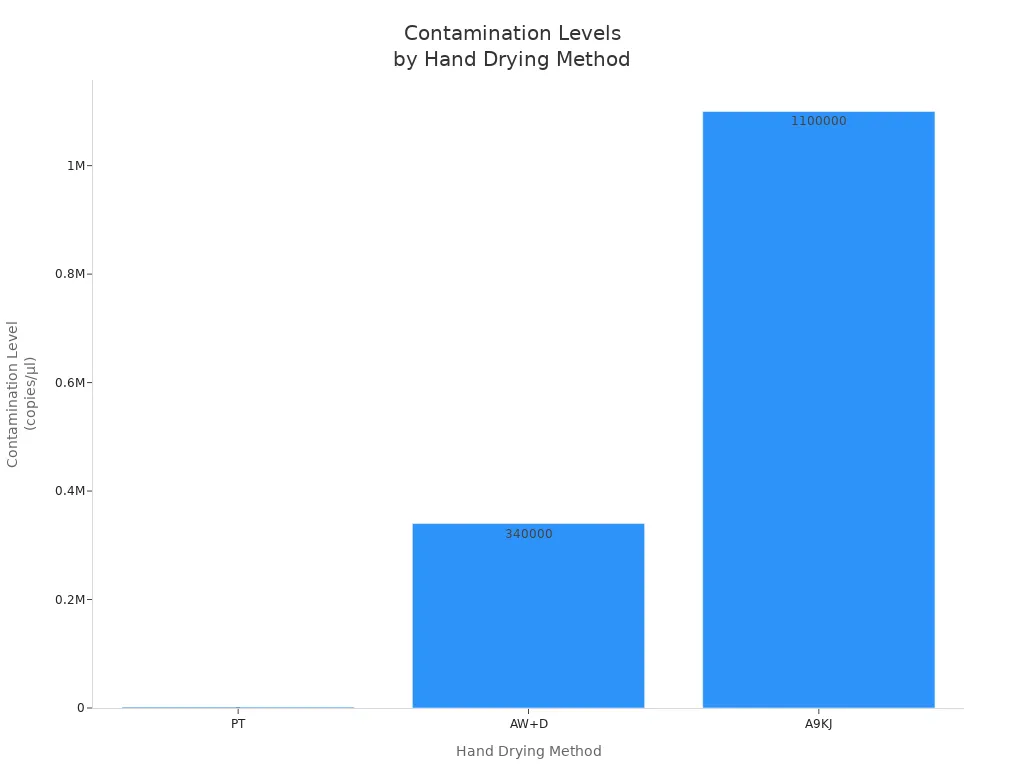
Hand drying with PT method leaves 200x fewer germs than AW+D (source) |
|
Soap Effectiveness |
Bar vs. liquid soap |
Liquid soap removes more bacteria |
Liquid soap reduces bacteria by up to 90% (CDC) |
|
Household Surfaces |
Sponge, faucet, remote |
Sponges show highest microbe count |
Kitchen sponges can hold up to 54 billion bacteria per cm² (National Geographic) |
|
Acid-Fast Stain Challenge |
Yogurt, sour cream |
Acid-fast bacteria stay red |
Acid-fast stain reveals Mycobacterium species (Microbiology Society) |
Note: Slides from science kits often have samples from things at home. You can use these to look at microbes and compare them with your own tests.
If you want to see how drying your hands changes germs, look at the chart above. The PT method leaves a lot fewer germs than AW+D or A9KJ. You can try this yourself with a microscope and slides. Test different ways to dry your hands and see which works best. You do not need fancy lab tools to explore the micro world. A simple microscope, a few slides, and curiosity help you discover new things. Each sample teaches you about hygiene, health, and the tiny world around you.
Why Viewing Germs Matters
Health and Hygiene Education
Looking at germs with a microscope helps you see why handwashing is important. You find out that many tiny living things are on things you touch. If you check your phone or a doorknob, you might see bacteria or fungi.
Sometimes, you spot something that looks like a tiny monster. Seeing germs makes hygiene feel real. You do not just hear about germs—you see them with your own eyes.
A microscope lets you test how well soap works. You can look at your hand before and after washing. You will see fewer germs after using soap. Try using the acid-fast stain to see which bacteria stay red. This shows which germs are harder to get rid of. With a compound microscope, you can see the difference between clean and dirty things. You learn that washing your hands helps keep you healthy.
Scientific Discovery and Research
You become a scientist when you use a microscope to study germs. When you look at a sample, you join a long line of discovery. Scientists first saw microorganisms with microscopes. They learned to tell different microbes apart and give them names. This helped create the germ theory of disease, which changed medicine. You can explore these ideas with your own microscope.
Here is a table that shows how microscopes helped science grow:
|
Discovery Type |
Description |
|---|---|
|
Observation |
You see microorganisms that were invisible before microscopes existed. |
|
Identification |
You can tell different microbes apart and learn their names. |
|
Germ Theory |
You understand that germs cause disease, thanks to what microscopes reveal. |
|
Research Advancement |
You help medicine, biology, and environmental science move forward. |
When you use a microscope, you do more than just look at germs. You ask questions, do experiments, and find answers. You might use a sample from yogurt, dirt, or pond water. Each time, you find something new. You learn how scientists use microscopes to fight sickness and keep people healthy. You see that research starts with a simple slide and a curious mind.
Step into a world of tiny wonders, invisible to the naked eye, where a microscope becomes your window to the unseen! With every glance through the lens, you uncover germs that influence your health and unlock secrets straight from science class. Imagine testing how thoroughly you wash your hands—or exploring everyday items up close in exciting home or classroom experiments. Each discovery sparks greater curiosity, and seeing those hidden microbes on your own skin brings home the importance of staying clean in a whole new way.
Get ready to explore, learn, and be amazed!
FAQ
Can you see all germs with a regular microscope?
You can see bacteria and fungi with a regular light microscope. Viruses are much smaller. You need an electron microscope to see them.
What is a prepared slide?
A prepared slide has a sample already fixed and stained. You just place it under your microscope and start exploring. Science kits often include these slides.
Why do you use stains like Gram stain?
Stains help you see germs better. Gram stain colors bacteria so you can tell different types apart. This makes it easier to study their shapes and features.
Is it safe to look at germs at home?
You stay safe if you use prepared slides or samples from clean surfaces. Always wash your hands after handling slides. Never use samples from dangerous places.

