Your skin holds countless secrets beneath its surface. With a microscope for skin, you can uncover intricate details about its structure and health that remain invisible to the naked eye. Skin conditions like atopic dermatitis, which affects up to 20% of infants globally, and skin cancer, with 3.6 million new cases annually in the USA, highlight the importance of understanding your skin at a deeper level.
Dermatology has adopted advanced microscopy to unveil hidden layers, detect early signs of disease, and examine the skin microbiome. These insights empower you to take control of your skin health like never before.
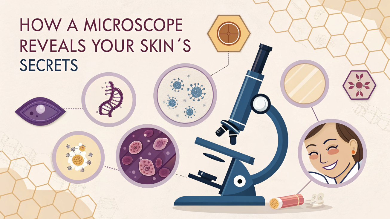
What Is a Microscope for Skin and How Does It Work?
Microscopes have changed the way you can explore the hidden world beneath your skin’s surface. A microscope for skin is a specialized tool designed to magnify and illuminate the intricate structures of your skin, revealing details invisible to the naked eye. These devices are not just ordinary microscopes; they are tailored to meet the unique challenges of skin analysis, offering unparalleled clarity and precision.
The Basics of Skin Microscopy
Skin microscopy utilizes advanced imaging techniques to examine the layers and components of the skin. Unlike standard microscopes, dermatology microscopes are equipped to handle the complex textures and varying depths of skin tissue. They allow you to observe everything from the outermost layer, the epidermis, to the deeper dermis, where blood vessels and collagen reside. Modern skin microscopes utilize different technologies to achieve high-resolution imaging.
For example, confocal laser scanning microscopy (CLSM) provides detailed, three-dimensional images of your skin’s microstructures. This technique uses laser light to focus on specific layers, enabling you to see cellular details without invasive procedures.
Researchers have found CLSM particularly useful for diagnosing conditions such as skin cancer and monitoring the effects of treatment.
Why Skin Analysis Requires Specialized Microscopes
Your skin is a dynamic organ with unique characteristics that demand specialized tools for accurate analysis. Dermatology microscopes are designed to address these needs. They offer features such as adjustable magnification, enhanced lighting, and the ability to distinguish between various skin components. These capabilities are essential for identifying subtle changes in skin texture, color, and structure.
Standard microscopes fall short when it comes to examining living skin. Specialized devices, such as the Dermascope Camera Probe, provide high-definition images that capture even the smallest details. These tools are invaluable for detecting early signs of conditions like atopic dermatitis or suspicious lesions.
Nonlinear optical microscopy, another advanced method, allows you to monitor treatment effects and assess the health of your skin’s layers without causing damage.
In dermatology, precision is key. High-resolution imaging tools like the Leica DM750 MOHS microscope enable dermatologists to differentiate between healthy and abnormal cells. This level of detail is crucial for early diagnosis and effective treatment planning. It will not matter if you’re exploring your skin’s microbiome or investigating a persistent rash; these microscopes empower you with insights that can transform your understanding of skin health.
Types of Microscopes for Skin Analysis
Understanding the different types of microscopes used in skin analysis can help you appreciate their unique capabilities. Each type serves a specific purpose, offering insights into your skin’s structure, health, and potential concerns.
Light Microscopes
Light microscopes are among the most commonly used tools in dermatology. These devices utilize visible light to magnify skin samples, enabling you to observe basic structures, such as cells and tissues. They are ideal for examining skin infections or infestations, as they provide clear and immediate results.
Direct microscopy, a technique often performed with light microscopes, is a simple and valuable tool for managing conditions such as fungal infections or scabies. While light microscopes are effective for basic analysis, their resolution is limited compared to more advanced options. They are most effective for surface-level observations and are commonly used in clinics for quick assessments. If you’re looking to explore deeper layers of the skin, other types of microscopes may be more suitable.
Confocal Microscopes
Confocal microscopes take skin analysis to the next level by providing high-resolution, three-dimensional images. These devices use a near-infrared laser to focus on specific layers of your skin, capturing intricate details of cellular structures like melanin, collagen, and keratin.
The pinhole mechanism ensures that only light from the focal plane reaches the detector, resulting in sharp and precise images. This technology is particularly useful for diagnosing inflammatory skin diseases, infections, and vascular abnormalities. Reflectance confocal microscopy (RCM), a specialized form of this technology, enables the examination of cellular morphology without the need for a biopsy. It offers unparalleled clarity, making it a valuable tool for evaluating skin diseases and monitoring cosmetic treatments like anti-aging creams or laser therapies.
Confocal microscopes also excel in noninvasive imaging. Nonlinear optical microscopy, for instance, enables you to visualize both the epidermal and dermal layers without causing any damage. This makes it an excellent choice for in vivo studies, where preserving the skin’s integrity is crucial.
Electron Microscopes
Electron microscopes offer the highest level of magnification and resolution, allowing you to see even the smallest details of your skin. Unlike light or confocal microscopes, these devices use a beam of electrons instead of light to create images. This enables them to reveal structures at the nanometer scale, such as individual collagen fibers or cellular organelles. These microscopes are invaluable for research and innovation in dermatology. They help scientists understand the microscopic changes associated with various skin conditions, paving the way for new treatments and technologies.
However, electron microscopes are not typically used for routine skin analysis due to their complexity and cost. They are best suited for advanced studies that require an in-depth examination of skin samples.
Dermatoscopes
Dermatoscopes are essential tools for examining your skin with precision. These handheld devices offer magnification ranging from 4x to 10x, enabling you to observe subsurface features that are invisible to the naked eye. Unlike traditional microscopes, dermatoscopes are specifically designed for dermatological purposes, making them invaluable for identifying skin abnormalities.
One of the key advantages of dermatoscopes is their ability to enhance clinical diagnosis. They help you detect subtle changes in skin lesions, such as variations in color, shape, or texture. This makes them particularly effective for spotting early signs of skin cancer, including melanoma. The DermaScope Camera Probe, a high-quality digital dermatoscope, takes this a step further by incorporating a polarized light source. This feature reduces surface reflections, giving you a clearer view of deeper skin structures.
Dermatoscopes are vital for tracking skin changes over time. Digital imaging with dermoscopy helps document and compare lesions at follow-ups, accurately monitoring progression or regression. Whether you’re a dermatologist or concerned about a mole, dermatoscopes offer detailed skin health insights.
What Microscopes Reveal About Your Skin
Skin Structure and Layers
Your skin is a complex organ composed of multiple layers, each serving a distinct purpose. A microscope for skin allows you to explore these layers in remarkable detail. The outermost layer, the epidermis, acts as a protective barrier against environmental damage. Beneath it lies the dermis, which contains essential components like collagen, elastin, and blood vessels that maintain your skin’s elasticity and nourishment.
At the deepest level, the hypodermis provides insulation and stores energy.
Dermatology microscopes reveal the intricate arrangement of cells within these layers. For example, confocal microscopes can highlight the distribution of melanin in the epidermis, enabling a better understanding of pigmentation patterns. Electron microscopes go even further, showing the nanostructures of collagen fibers that contribute to your skin’s firmness. These insights are invaluable for understanding how your skin functions and how it responds to aging, environmental stress, or skincare products.
Skin Conditions and Concerns
Microscopes play a crucial role in identifying and understanding various skin conditions. Many issues, such as acne, eczema, and psoriasis, originate at the cellular level. Dermatology microscopes allow you to observe these conditions closely, providing a clearer picture of their causes and progression. For instance, light microscopes can detect fungal infections by revealing the presence of fungal hyphae in skin samples. Confocal microscopes, on the other hand, can identify inflammatory markers in conditions like rosacea or dermatitis.
Skin cancers, one of the most serious concerns, can also be detected early with the help of advanced microscopy. Dermatoscopes are particularly effective for examining suspicious moles or lesions. They magnify subsurface features, making it easier to spot irregularities in color, shape, or texture.
Reflectance confocal microscopy provides a non-invasive method for analyzing cellular structures, thereby reducing the need for biopsies. Early detection through these tools significantly improves treatment outcomes.
Microscopes also aid in monitoring the effectiveness of treatments. Whether you’re using topical creams for acne or undergoing laser therapy for pigmentation, these devices help track changes in your skin over time. This ensures that your treatment plan is working as intended and allows for adjustments if necessary.
Microbiome and Skin Health
Your skin is home to a diverse community of microorganisms, collectively known as the skin microbiome. These microbes play a vital role in maintaining your skin’s health. Some protect against harmful pathogens, while others contribute to processes like wound healing.
Microscopes provide a window into this microscopic world, helping you understand how these organisms interact with your skin.
Studies have shown that the composition of your skin microbiome varies across different areas of your body. For example, metagenomic analysis has revealed distinct microbial communities at 18 different skin sites. Microscopes enable researchers to study these variations, shedding light on how they affect skin conditions such as acne or eczema. Swab and biopsy methods are commonly used to collect samples for such studies, with each method offering unique advantages.
The role of the microbiome in wound healing is another area of interest. Certain skin commensal organisms have been found to accelerate the healing process, while others may hinder it. Microscopes help identify these beneficial and harmful microbes, paving the way for targeted treatments.
Understanding your microbiome can also help you choose skincare products that support a healthy microbial balance. Microscopic insights into the microbiome are not just for researchers. They empower you to make informed decisions about your skincare routine. For instance, products containing probiotics or prebiotics can promote a balanced microbiome, enhancing your skin’s natural defenses.
With the help of microscopy, you can take a proactive approach to maintaining your skin’s health and resilience.
Gaining microscopic insights empowers your skincare approach. Blending professional guidance with a personalized routine allows you to tackle current issues and prevent future problems. This proactive method helps keep your skin healthy, durable, and glowing.
Microscopes for skin offer a groundbreaking way to understand your skin’s health. These tools enable you to detect issues early, such as skin cancer or infections, and guide personalized care plans. Advances in dermatology, like confocal microscopy and dermoscopy, have made noninvasive diagnostics more accurate and accessible.

