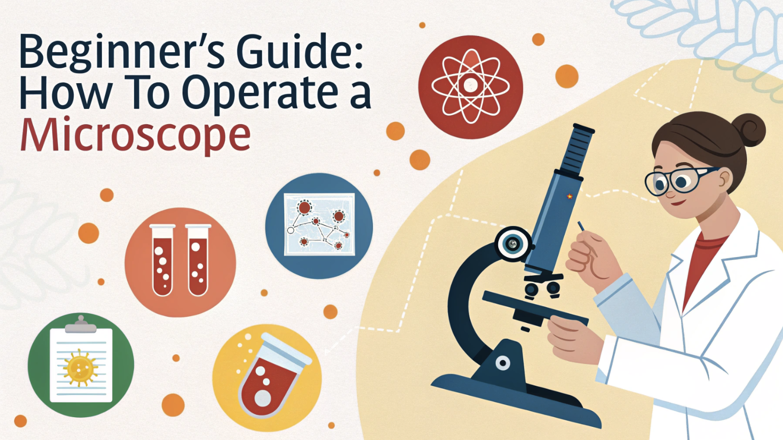
So, you’ve just got your hands on a microscope, huh? Exciting stuff! Maybe it’s for a school project, or you’re just curious about the tiny world around us. Either way, you’re probably staring at all those knobs and lenses thinking, “Uh… now what?” Don’t sweat it – we’ve all been there!
Remember when you first learned to ride a bike? This is kind of like that, but instead of skinned knees, you might end up with a sore eye from squinting. But don’t worry! This beginner’s guide will walk you through the basics of operating a microscope, step by step. We’ll cover everything from turning it on (yes, that’s actually a step) to focusing on your first specimen. Let’s get started, shall we? 🙌🏼
Understanding Microscope Basics
Before going into the specifics of operating a microscope, it’s important to understand its basic components. In this section, we will explore the anatomy of a microscope, including the eyepiece, objective lenses, stage, and focus knobs.
Understanding these fundamentals will lay the foundation for operating the microscope effectively.
Anatomy of a Microscope
Let’s familiarize ourselves with the key parts of this fascinating instrument:
- Eyepiece: The eyepiece, also known as the ocular lens, is where you place your eyes to view the specimen. It typically provides a magnification of 10x.
- Objective Lenses: The objective lenses are located on a revolving nosepiece below the eyepiece. They come in different magnifications, such as 4x, 10x, 40x, and 100x, allowing you to observe the specimen at various levels of detail.
- Stage: The stage is a platform that holds the specimen slide in place. It usually has a mechanical stage with knobs for precise movement of the slide.
- Focus Knobs: The focus knobs consist of the coarse adjustment knob and the fine adjustment knob. The coarse knob moves the stage rapidly, while the fine knob allows for delicate adjustments to achieve sharp focus.
Microscope Components
Optical Components
Let’s start with the heart of your microscope: the optical components. These are the parts that do the magnifying and trust me, they’re pretty cool once you get to know them.
First up, we have the eyepiece, also known as the ocular lens. This is where you’ll press your eye to peer into the microscopic world. Most microscopes have eyepieces that magnify 10x, but you can find some that go up to 15x or even 20x.
Pro tip: if you wear glasses, look for microscopes with high eye-point eyepieces. These allow you to keep your glasses on while using the microscope, saving you from the awkward dance of taking your glasses on and off.
Next, let’s talk about the star of the show: the objective lenses. These bad boys do the heavy lifting when it comes to magnification. You’ll usually find a set of three or four objectives on a rotating nosepiece. Each objective has a different magnification power, typically ranging from 4x to 100x. When you’re just starting, stick with the lower magnifications. It’s easier to find your specimen and get it in focus. As you become more comfortable, you can move up to the higher powers for more detailed viewing of the specimens.
Here’s a quick reference table for typical objective lens magnifications:
| Objective | Magnification | Common Uses |
|---|---|---|
| Scanning | 4x | Locating specimens, overview |
| Low Power | 10x | General viewing, larger structures |
| High Power | 40x | Cellular details, smaller organisms |
| Oil Immersion | 100x | Bacteria, cell organelles |
Moving down the microscope, we come to the stage and stage clips. This is where you’ll place your slides. The stage clips hold your slide in place, which is crucial for keeping your specimen in view as you focus. Some high-end microscopes feature mechanical stages that enable precise movement of the slide using knobs.
If you’re serious about microscopy, this feature is a game-changer. Underneath the stage, you’ll find the condenser and diaphragm (or iris). These components control the light that illuminates your specimen. The condenser focuses the light onto your specimen, while the diaphragm controls how much light gets through. Proper use of these can dramatically improve the contrast and clarity of your image.
Remember, more light isn’t always better – sometimes reducing the light can reveal details you might otherwise miss.
Mechanical Components
Now, let’s get into the nuts and bolts of your microscope – literally! The mechanical components are what hold everything together and allow you to make those crucial adjustments.
The base and arm of the microscope might seem boring, but they’re essential. The base provides stability, preventing those frustrating moments when your microscope wobbles just as you’re about to make a crucial observation. The arm allows you to carry the microscope safely; always use two hands, one on the arm and one to support the base. Some microscopes have tilting heads, which can be a lifesaver for your neck during long observation sessions.
Next up are the focus knobs – your best friends in the quest for crystal-clear images. You’ll typically find two types: coarse and fine focus knobs. The coarse focus knob allows for large adjustments, helping you roughly bring your specimen into focus. Once you’re close, switch to the fine focus knob for those minute adjustments that bring everything into sharp relief.
Here’s a tip: always start with your objective lens close to the slide and focus upwards. This prevents you from accidentally crushing your slide (and potentially your objective lens) by focusing downwards too far.
Lastly, we have the revolving nosepiece. This is the circular part that holds your objective lenses. It allows you to switch between different magnifications quickly and easily. Always make sure you hear a ‘click’ when rotating to a new objective – this ensures it’s properly aligned. And here’s a cool trick: if you’re having trouble finding your specimen under high magnification, switch to lower power, center your specimen, and then rotate back to high power. It’ll save you a lot of frustration!
Remember, getting to know these components isn’t just about memorizing names. It’s about understanding how each part contributes to revealing the hidden world beneath your microscope. The more familiar you become with these components, the more you’ll be able to coax out of your microscope, revealing details and structures you might have missed before.
Preparing to Use a Microscope
Setting Up Your Workspace
Your workspace can make or break your microscopy experience, so let’s get it right from the start.
First, choose a location that’s stable and vibration-free. That wobbly desk in the corner?
Not ideal.
Vibrations are the enemy of clear images, so aim for a solid, sturdy surface. If you’re dealing with an unavoidably shaky spot, try placing a rubber mat under your microscope to absorb some of the vibrations. Lighting is crucial in microscopy, but it’s often overlooked. Natural light can be great, but it’s inconsistent. Instead, opt for a dedicated microscope lamp or a good desk lamp. Position it so it doesn’t cast shadows on your work area.
Here’s a pro tip: if you’re using a separate lamp, place it slightly to the left if you’re right-handed (or right if you’re left-handed). This setup prevents your hand from casting shadows as you manipulate slides. Comfort is key for long observation sessions. Adjust your chair height so your eyes are level with the eyepieces when sitting up straight. This prevents neck strain and makes it easier to maintain proper posture.
Keep your tools—slides, cover slips, and lens paper—within easy reach.
A clean, organized workspace isn’t just satisfying; it’s efficient.
Cleaning and Maintenance

Now, let’s discuss maintaining your microscope in optimal condition. A clean microscope isn’t just more pleasant to use; it provides clearer, more accurate images. Plus, proper maintenance extends the life of your instrument. Start with the lenses – they’re the most critical and delicate parts of your microscope. Use only lens paper or a microfiber cloth designed for optics. Never use regular tissues or your shirt – they can scratch the lens!
For stubborn smudges, breathe gently on the lens to fog it up, then wipe in a circular motion. Avoid using cleaning solutions unless necessary, and then only use those specifically designed for optical lenses.
Here’s a cleaning schedule to keep your microscope in prime condition:
| Component | Cleaning Frequency | Method |
|---|---|---|
| Eyepieces | After each use | Lens paper, gentle circular motion |
| Objectives | After each use | Lens paper, gentle circular motion |
| Stage | Daily | Soft brush, then damp cloth |
| Body | Weekly | Soft cloth, mild soap if needed |
Don’t forget about the mechanical parts. A tiny drop of microscope-grade oil on the focus mechanisms every few months can keep things running smoothly. However, be cautious – excessive oil can attract dust and grime.
Storage is an often-overlooked aspect of microscope care. Always cover your microscope when not in use to protect it from dust. If you’re storing it for an extended period, remove the eyepieces and objectives and store them separately in a dry, dust-free container. This prevents moisture from getting trapped between the lenses, which can lead to fungal growth – a microscope’s worst enemy. Remember, a well-maintained microscope is a joy to use. It provides clearer images, operates more smoothly, and lasts longer.
Basic Microscope Operation
Powering On and Initial Setup
Alright, let’s get this show on the road! Powering on your microscope might seem straightforward, but there’s a method to the madness that’ll save you time and frustration.
First things first, always start with the lowest magnification objective lens. This gives you the widest field of view, making it easier to locate your specimen. It’s like using a map before zooming in on Google Earth – you want to know where you are before diving into the details.
Next, adjust your light source. If you’re using a microscope with a built-in illuminator, start with it on the lowest setting. You can always increase the brightness, but starting too bright can be harsh on your eyes and wash out details in your specimen. For microscopes with a mirror, angle it to reflect ambient light up through the stage.
Now, let’s talk about those eyepieces. If your microscope has adjustable eyepieces, take a moment to set them correctly. Close one eye and adjust the eyepiece for the open eye until the image is sharp. Repeat for the other eye. This step is crucial for comfortable viewing, especially during long observation sessions.
Remember, your eyes are unique – don’t be afraid to fine-tune these settings for your personal comfort.
Preparing and Mounting Specimens
Creating the perfect slide is an art form in itself. Whether you’re using a prepared slide or creating your own, proper technique is essential for clear, detailed observations. For wet mounts, less is more. Place a small drop of water or staining solution on your slide, then gently lower the coverslip at an angle. This prevents air bubbles, which can obstruct your view. If you do get bubbles, try gently tapping the coverslip with a pencil eraser – it often helps to dislodge them.
When using prepared slides, always handle them by the edges to avoid smudging. Before placing the slide on the stage, give it a quick once-over with a lint-free cloth. It’s amazing how often a tiny smudge or piece of dust can be mistaken for part of the specimen!
Here’s a quick reference for common staining techniques:
| Stain | Color | Best for |
|---|---|---|
| Methylene Blue | Blue | Bacteria, cell nuclei |
| Iodine | Brown | Starch in plant cells |
| Eosin | Pink/Red | Cytoplasm, red blood cells |
| Crystal Violet | Purple | Gram staining bacteria |
Remember, different stains highlight different structures. Experimenting with various stains can reveal new aspects of your specimens. Just be sure to label your slides accurately – future you will thank present you for the clear notes!
Focusing and Magnification
Now for the moment of truth – bringing your specimen into focus. This is where patience pays off, folks. Rush this step, and you might miss the best parts of your sample.
Start with your coarse focus knob. Look at the stage from the side and slowly raise the objective until it’s close to the slide – but not touching! Now, peer through the eyepiece and slowly lower the objective using the coarse focus. Stop as soon as you see your specimen come into view. Once you’ve got a rough focus, switch to the fine focus knob. This is where the magic happens. Make tiny adjustments until your image snaps into crystal-clear focus.
If you’re having trouble, try this trick: slightly close the diaphragm to increase contrast. It can make focusing easier, especially on nearly transparent specimens.
Ready to zoom in? Here’s how to change magnification like a pro:
- Center your specimen in the field of view
- Rotate the nosepiece to the next highest objective
- Use only the fine focus to sharpen the image
- Adjust the light intensity if needed
Do note that as you increase magnification, you decrease your field of view and depth of field. This table shows typical relationships:
| Objective | Total Magnification* | Field of View | Depth of Field |
|---|---|---|---|
| 4x | 40x | Large | Deep |
| 10x | 100x | Medium | Moderate |
| 40x | 400x | Small | Shallow |
| 100x | 1000x | Very Small | Very Shallow |
*Assuming a 10x eyepiece
As you move to higher magnifications, you’ll need to make more frequent fine focus adjustments. This is because the depth of field becomes shallower, meaning less of your specimen is in focus at any given time.
It’s like switching from a landscape photo to a macro shot – you’ve got to be more precise with your focus.
One last pro tip: if you lose your specimen at high magnification, don’t panic! Simply lower the magnification, re-center your specimen, and then increase the magnification again. It’s a common issue even for experienced microscopists, so don’t get discouraged if it happens to you.
Advanced Microscope Techniques
Using Oil Immersion
Ready to take your microscopy skills to the next level? Let’s get into oil immersion. This technique enables the use of ultra-high magnification objectives (typically 100x) by eliminating the air gap between the specimen and the lens.
First, you’ll need immersion oil. This isn’t just any oil – it’s specially formulated to match the refractive index of glass. Start by focusing on your specimen using your 40x objective. Once you’ve got a clear image, rotate the nosepiece halfway between the 40x and 100x objectives. Place a small drop of oil on the slide, right over your specimen.
Now, carefully rotate the 100x objective into place.
Here’s where things get tricky. You won’t be able to see anything at first, and you can’t use the coarse focus at this magnification. Instead, use the fine focus knob to slowly bring your specimen into view. It might take some practice, but the results are worth it. You’ll be able to see incredible detail, like the individual flagella on bacteria or the intricate structures within plant cells.
Remember, oil immersion isn’t just a cool trick – it’s essential for certain types of microscopy. For example, proper identification of many bacterial species requires oil immersion to see key diagnostic features.
Just don’t forget to clean your objective thoroughly after use. Oil left on the lens can harden and damage it over time.
Adjusting Illumination
Mastering illumination is like learning to paint with light. It can dramatically improve the contrast and clarity of your images, revealing details you might otherwise miss. Let’s start with the condenser. This often-overlooked component focuses light onto your specimen. For most observations, you’ll want the condenser as close to the stage as possible. Use the condenser focus knob to adjust it until your field of view is evenly illuminated.
Next up is the diaphragm or iris. This controls the amount of light that reaches your specimen. Counter-intuitively, more light isn’t always better. Closing the diaphragm increases contrast, which can be crucial for seeing nearly transparent specimens.
Here’s a general rule of thumb:
| Magnification | Diaphragm Opening |
|---|---|
| Low (4x-10x) | Wide open |
| Medium (40x) | Partially closed |
| High (100x) | Nearly closed |
But don’t just set it and forget it. Experiment with different diaphragm settings to find what works best for each specimen. You might be surprised at how much detail you can reveal with the right illumination.
For the true illumination aficionados, there’s Köhler illumination. This technique ensures optimal and even illumination of your specimen. It involves aligning and focusing your light source, condenser, and objective to create a perfectly even field of illumination. It takes some practice to master, but once you’ve got it down, you’ll never want to go back to standard illumination.
Remember, proper illumination isn’t just about seeing your specimen clearly. It also helps prevent eye strain during long observation sessions. Your future self will thank you for taking the time to get it right!
Troubleshooting Common Issues
Image Quality Issues
Let’s face it, even the most seasoned microscope users occasionally encounter image quality issues. But don’t worry, most problems have simple solutions once you know what to look for.
Blurry images are probably the most common complaint. If your specimen looks like it’s underwater, first check your focus. Are you using the fine focus knob for higher magnifications? Remember, as magnification increases, your depth of field decreases, making precise focus crucial. If you’re sure your focus is spot-on, check your lenses for dirt or smudges.
A tiny fingerprint can cause a major blur. Clean your lenses gently with lens paper, using a circular motion from the center outward.
Poor contrast can make details hard to see, especially with nearly transparent specimens. Try adjusting your illumination. Close down your diaphragm a bit to increase contrast. If that doesn’t help, consider using a stain appropriate for your specimen. Here’s a quick guide to some common stains and their effects:
| Stain | Color | Enhances |
|---|---|---|
| Methylene Blue | Blue | Cell walls, nuclei |
| Iodine | Brown | Starch granules |
| Safranin | Red | Cell walls, chromosomes |
| Nigrosin | Black | Background (negative stain) |
Sometimes, you might notice strange artifacts in your image – things that clearly aren’t part of your specimen. Before you get excited about discovering a new microorganism, check for air bubbles or debris on your slide.
Air bubbles look like perfect circles with dark edges. To avoid them, lower your coverslip at an angle when preparing slides.
For debris, make sure you’re using clean slides and coverslips, and keep your work area dust-free.
Mechanical Problems
Mechanical issues can be frustrating, but many are easy to fix with a little know-how.
Let’s tackle some common problems.
If your focus knobs are sticking or difficult to turn, avoid forcing them. This usually indicates a lubrication issue. First, try gently cleaning the focus mechanism with a soft, lint-free cloth. If that doesn’t help, you might need to apply a tiny amount of microscope-grade lubricant.
Be careful though – too much oil can attract dust and make the problem worse. Here’s a quick guide to lubricating different parts:
| Component | Lubricant Type | Application Frequency |
|---|---|---|
| Focus Knobs | Light machine oil | Every 6 months |
| Stage | Dry lubricant | Annually |
| Nosepiece | Grease | Every 2 years |
A misaligned nosepiece can cause your objectives to be off-center, making it hard to keep your specimen in view when changing magnification. First, check if your nosepiece is loose. Many microscopes have a small set screw that can tighten the nosepiece. If tightening doesn’t solve the problem, you might need to realign your objectives. This is a delicate process best left to professionals, but here’s a quick test: switch between objectives and see if your specimen stays centered. If not, it’s time for a tune-up.
Stage drift is another common annoyance. You focus on a perfect specimen, look away for a second, and suddenly it’s gone! This usually happens when your stage isn’t quite level. Check if your microscope has leveling screws on the base. If so, use a small bubble level to ensure your microscope is perfectly horizontal. No leveling screws? Try placing thin shims under the microscope base to level it out.
Remember, while many mechanical issues can be resolved with simple fixes, don’t hesitate to seek professional help for more serious problems. A well-maintained microscope will provide years of crystal-clear observations, so it’s worth investing in proper care and occasional professional servicing.
Summing Up
We’ve covered a lot of ground, from the basics of focusing to some advanced techniques that are pretty cool. But here’s the thing – operating a microscope is a skill you’ll keep developing. Don’t sweat it if you’re not perfect right away. The key? Start with the basics: clean equipment, careful preparation, and lots of practice. As you become more comfortable, experiment with various techniques. Each specimen has its own secrets, and half the fun is figuring out how to reveal them.
Remember, microscopy is as much about curiosity as it is about technical skill. Continue asking questions, take notes, and share your findings. There’s a whole tiny world out there waiting for you to explore.

