Light microscopes have been an important part of scientific advancement since the 4th century AD till today. They operate by bending light, using refraction to create magnified images. Different types of light microscopy, such as brightfield and darkfield, are utilized for various purposes. Magnifications of up to 1,000x can be achieved with compound microscopes.
Major brands like Nikon, Olympus, Leica, Zeiss, and Motic offer high-quality light microscopes for diverse applications.
- Introduction to Light Microscopy
- 🔊 Play Audio Version
- Basic Components of a Light Microscope
- How Do Light Microscopes Work
- Step-by-Step Guide on Using a Light Microscope
- Types of Light Microscopy Techniques
- 10 Modern Applications of Light Microscopes
- Advanced Light Microscopy Methods
- Popular Light Microscope Brands
- To Sum Up
Introduction to Light Microscopy
Light microscopy started in the 4th century AD. It uses visible light to make small objects appear larger. This technology is key in biology, nanotechnology, and forensics today.
What is Light Microscopy?
It uses glass lenses to focus light. This lets us see very tiny things. There are simple and compound microscopes that magnify objects. In labs, various methods are used to visualize objects clearly, such as brightfield and phase contrast microscopy.
Applications of Light Microscopy
In medicine, it aids in diagnosing diseases. It’s also vital in studying cells and tissues in the biological sciences.
In materials science, it examines the structures of new materials. Also, it plays an instrumental role in verifying product quality, conducting surface checks, and facilitating crime scene investigations. Light microscopes also help students learn about cells and biology.
Recent advances allow for even more detailed views of small structures. This is useful in nanotechnology for studying and working with tiny structures.
Brands |
Specialization |
|---|---|
| Nikon | High-precision research microscopes |
| Olympus | Educational and clinical microscopes |
| Leica Microsystems | Innovative optical instruments |
| Zeiss | Advanced imaging systems |
| Motic | Cost-effective educational microscopes |
🔊 Play Audio Version
Basic Components of a Light Microscope
In this section, we will examine the key components of a light microscope closely. We’ll learn how these parts work together to help us see small details more clearly.
Illumination System
The illumination system lights up the sample. It often uses LED or halogen lights to ensure the light remains consistent. Special methods using light can reveal how crystals and minerals are structured.
Imaging System
The imaging system makes the tiny thing we are looking at appear big. It has lenses that zoom in on the sample. Sometimes it uses filters to make the image clearer. This part is important for magnifying details.
Objective Lenses
Objective lenses are key parts. They help gather light from the sample and make it bigger and clearer. By using multiple lenses, we can zoom in a lot closer. They can make things seem 4 to 100 times larger. This depends on the microscope.
Eyepieces
Eyepieces make the already enlarged image even bigger for our eyes. They are crucial and add to how much we can see. They usually make things 8 to 12 times larger. The final zoom level comes from the eyepieces and the objective lenses working together.
Component |
Function |
Popular Brands |
|---|---|---|
| Illumination System | Provides light to the sample | Nikon, Olympus, Leica Microsystems, Zeiss, Motic |
| Imaging System | Forms the magnified image | Nikon, Olympus, Leica Microsystems, Zeiss, Motic |
| Objective Lenses | Collect and magnify light from the sample | Nikon, Olympus, Leica Microsystems, Zeiss, Motic |
| Eyepieces | Further enlarge the image for observation | Nikon, Olympus, Leica Microsystems, Zeiss, Motic |
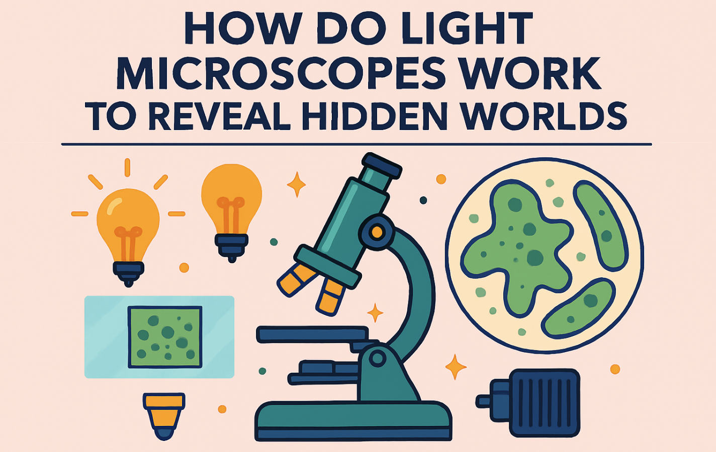
How Do Light Microscopes Work
Figuring out light microscopes is like solving a science mystery. They work by utilizing the concept of light refraction, which involves bending light as it passes through different materials, allowing us to see larger images of smaller things. Now, let’s look closer at this cool trick.
Principles of Light Refraction
Light microscopes are based on the rule of light bending. This happens when light goes from one thing to another, like air to glass. When this happens, the light changes speed and direction because of the material’s refractive index. The lenses in microscopes have a special shape that focuses light, making objects appear larger.
This is similar to how our eyes work with lenses to focus objects on the retina, the back wall of the eye.
Image Formation and Magnification
Different parts have to work well together for a light microscope to produce clear images, Things get bigger in the microscope mainly because of the objective lens and the eyepiece. The objective lens can magnify objects from 4 to 100 times, and the eyepiece adds another 10 times. Besides making things look bigger, microscopes have knobs to adjust focus and various special settings to enhance the visibility of tiny details.
When light goes through the microscope, colors and varieties of light help us see things clearly. They help us study tiny life forms and materials better by using light bending.
Step-by-Step Guide on Using a Light Microscope
Using a light microscope properly not only enhances the accuracy of your observations but also extends the lifespan of this precise instrument. Whether you’re a student, hobbyist, or professional, following a systematic approach will help you achieve the best results.
Here’s a straightforward guide to help you get started:
Preparing Slides for Viewing
To begin, you’ll need a well-prepared slide. Here’s how you can prepare one:
- Clean the Slide: Always start with a clean slide and cover slip. Any dust or smudges can obscure your view.
- Place the Specimen: Place your specimen on the center of the slide. If it’s a liquid sample, just a drop is sufficient.
- Cover the Specimen: Gently lower a cover slip over the specimen to avoid trapping air bubbles.
Setting Up the Microscope
Setting up your microscope correctly is crucial for optimal viewing:
- Turn on the Illumination: Ensure that your light source is on and functioning.
- Start with the Lowest Magnification: Place the objective lens with the lowest magnification (usually 4x or 10x) into position.
- Adjust the Eyepiece: If your microscope has an adjustable eyepiece, set it to match your eyesight.
Adjusting Focus and Magnification
Focusing on your specimen is perhaps the most critical step in using a microscope:
- Coarse Adjustment: Use the coarse focus knob to move the stage up, bringing the specimen closer to the objective lens. Look through the eyepiece and adjust until the image comes roughly into focus.
- Fine Adjustment: Switch to the fine focus knob to sharpen the image.
- Increase Magnification: Once focused, you may switch to a higher magnification if needed. Re-focus using the fine adjustment knob each time you switch.
Tips for Effective Microscope Use
To maximize your microscopy experience, consider these tips:
- Always Start with Low Power: This makes it easier to locate your specimen and reduces the risk of damage.
- Use Both Eyes: Try to keep both eyes open when looking through a monocular microscope to avoid eye strain. =json
- Adjust the Light: Use the iris diaphragm to adjust the amount of light passing through the specimen for clearer images.
Common Adjustments and Their Effects
| Adjustment | Function | Expected Effect |
|---|---|---|
| Coarse Focus | Raises/lowers the stage quickly. | Brings specimen into general focus. |
| Fine Focus | Raises/lowers the stage slowly. | Sharpens the image. |
| Iris Diaphragm | Controls light intensity. | Enhances contrast and detail. |
| Objective Lens | Changes magnification. | Increases detail as magnification increases. |
Using a light microscope effectively involves understanding how each component affects your viewing experience. By following these steps, you can ensure clear, detailed observations and take proper care of your equipment.
Types of Light Microscopy Techniques
It’s key to understanding different light microscopy approaches. These methods utilize light to reveal tiny details that would otherwise be invisible.
Brightfield Microscopy
Brightfield microscopy is very common. It uses halogen lamps and LED lights for clear views. However, it needs samples to be stained for better contrast. It’s used a lot in schools and labs.
Darkfield Microscopy
Darkfield microscopy makes a dark background, helping see edges better. It’s great for watching cells without staining. It uses a special setup to create a bright image on a dark field.
Phase Contrast Microscopy
Phase contrast microscopy is well-suited for examining transparent samples. It changes light shifts into intensity changes. This makes it ideal for viewing cells and structures clearly, without the need for staining.
Brightfield, Darkfield, and Phase Contrast are helpful tools. They allow scientists to choose the best method for studying their subject. This enhances our ability to see and understand very small things.
10 Modern Applications of Light Microscopes
Medical Diagnosis
Light microscopes are crucial in pathology for examining tissue samples to diagnose diseases such as cancer. They enable pathologists to observe the morphology of cells and tissues in detail.
This microscopic analysis helps identify abnormal cell structures characteristic of various cancers and other diseases, enabling accurate diagnoses and treatment plans.
Microbiology
In microbiology, light microscopes are used to observe bacteria, viruses, and other microorganisms. This helps in studying their structure, behavior, and in identifying new species or pathogens. Microscopes facilitate the visual examination of organism shapes, sizes, and mobilities, which are critical for classifying and understanding their roles in different environments.
Education
Light microscopes are fundamental tools in educational institutions for teaching biology. They allow students to explore cell structures, microorganisms, and biological processes, enhancing their understanding of the microscopic world. Through direct observation, students can appreciate the complexity of life at a cellular level, fostering a deeper interest and knowledge in biological sciences.
Environmental Science
Researchers use light microscopes to study environmental samples, such as water and soil, to monitor pollutants and assess the ecological health of these environments. This microscopic analysis helps identify contaminants, such as microplastics, pathogens, and other particulates, that can impact ecosystems.

Pharmacology
In drug development, light microscopes help in the examination of cellular responses to new pharmaceuticals. This aids in understanding drug efficacy and safety by observing how cells react to treatments at a microscopic level, which is crucial for developing effective and safe medications.
Food Industry
Microscopes are used in the food industry to ensure quality control by detecting foreign particles and microorganisms in products. This microscopic examination helps maintain food safety standards and prevent contamination that could lead to foodborne illnesses.
Forensic Science
Light microscopes play a role in forensics for analyzing samples such as fibers, hair, or blood traces, which can be pivotal in solving crimes. The detailed visual information obtained can link suspects to crime scenes or victims, providing critical evidence in legal cases.
Materials Science
They are employed to examine the structure and properties of materials, such as metals, polymers, and ceramics, to enhance their performance and durability. Microscopes enable scientists to study material grains, phases, and defects at a microscopic level, informing enhancements in engineering and manufacturing processes.
Botany
Light microscopes facilitate the study of plant cells and tissues, enabling botanists to understand plant anatomy and pathology, which is crucial in agriculture and horticulture.
Microscopic analysis helps in identifying disease patterns, understanding plant development, and in breeding more robust plant varieties.
Genetics
Microscopes are used in genetics for chromosome mapping and identifying genetic disorders. They help in visualizing structures within a cell that are crucial for genetic research and diagnostics. This includes observing the arrangement and number of chromosomes, identifying genetic mutations, and examining gene expression at a cellular level, which can lead to breakthroughs in medical treatments and a deeper understanding of hereditary diseases.
Advanced Light Microscopy Methods
When we discuss sophisticated microscopy, fluorescence microscopy, and confocal microscopy take center stage. They provide exceptionally clear visuals. These techniques are invaluable in fields such as cell biology, and immunology, among many others.
Fluorescence Microscopy
Fluorescence microscopy uses a special light to make things glow. It shows us tiny parts in cells and biology very clearly. This method is key in studying cells, immune responses, and details of molecules.
It also lets us see cell parts by using light and specific antibodies. These make details pop out in color.
Confocal Microscopy
Confocal microscopy goes a step further. It uses a laser and a special lens to see things clearly. This way, researchers can look at cells and their actions in 3D.
This method is ideal for capturing clear images of small cell components. It helps in biology studies a lot.
Both types of microscopy are key in today’s science. They let us see the world of biology in ways we never could before. This helps us make new, important discoveries.
Popular Light Microscope Brands
Several top brands are leading in high-quality microscopes. They are known for their precision, innovation, and design. Let’s explore the best-known names in detail.
Nikon Microscopes
Nikon leads with its top-notch optics and sturdy construction. Their microscopes are perfect for research and learning. They offer clear, precise images. Nikon excels in innovation, winning over many pros.
Olympus Microscopes
Olympus is a big name in microscopes, with a focus on quality optics for research, health, and school. Their microscopes shine with sharp imaging and easy use. They’re great for busy labs. Olympus stands out for being easy to use and upgrade for various microscopy techniques including Differential Interference Contrast (DIC).
Leica Microsystems
Leica is known for advanced microscopes you can customize. Their software and accessories help you work better, especially in research. Leica makes sure your images are accurate, with less risk of injury when working long hours.
Zeiss Microscopes
Zeiss is famous for its advanced optics and technology. Their microscopes offer clear, detailed images for research and science. Zeiss microscopes can be customized and have great service. Buyers love their strong image capabilities and flexible design.
Motic Microscopes
Motic is known for making quality microscopes affordable. They’re great for schools and beginners. Motic microscopes find the right balance between cost and quality. Though they may not offer as many upgrades, Motic meets basic microscope needs well.
To Sum Up
Exploring the tiny world through a microscope is like a beautiful show. It shows us things we can’t see with our eyes alone. Due to the invention of light microscopes, it is possible to learn more about tiny cells and creatures. They are a key tool for scientists in various fields, including biology, materials science, and even crime investigation.

