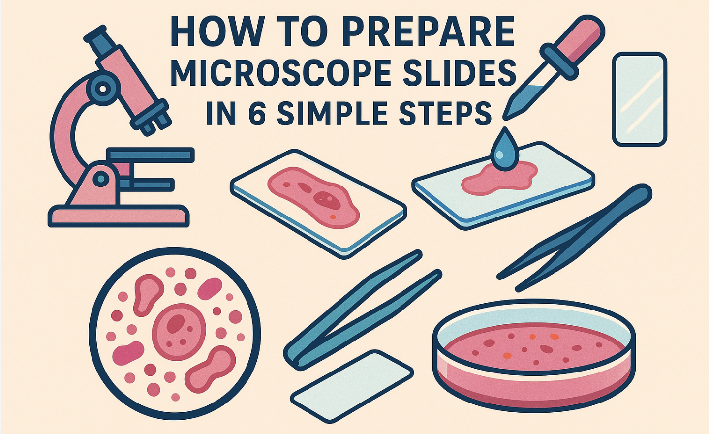
Imagine peering into a world invisible to the naked eye, where every detail matters. That’s the magic of microscope slides. Preparing them correctly is crucial. A single error can lead to misdiagnosis or incorrect treatment.
In fact, poorly prepared slides can result in significant clinical impacts, affecting up to 60-70% of medical decisions.
Microscope slides are not just tools; they are gateways to discovery. They allow us to explore the intricate details of specimens, providing insights that drive scientific research forward. Knowing how to prepare microscope slides properly ensures accuracy and reliability, making every observation count.
Step 1: Gather Necessary Tools and Materials
To get started with slide preparation, it’s essential to have all the necessary tools and materials within reach. Gathering everything you need beforehand helps ensure a smooth and efficient process. This preparation stage is crucial for a successful microscopic examination.
Tools Required
-
Microscope slides: These transparent glass or plastic pieces are the foundation of your observation. They support the specimen, allowing light to pass through for clear viewing.
-
Cover slips: These thin glass pieces protect the specimen and help flatten it for better focus.
-
Specimen samples: Choose your sample wisely. Whether it’s a plant leaf or a drop of pond water, the specimen is the star of the show.
-
Staining solutions: These enhance contrast, making cell structures more visible. Choose a stain that suits your specimen type.
-
Droppers and pipettes: These tools help you apply liquids precisely, ensuring no mess and no fuss.
Supporting Stats
Using clean slides is crucial for accurate results. A study found that contaminated slides can lead to errors in up to 30% of observations. Clean slides ensure clarity and precision, making every detail count.
Common Mistakes to Avoid
One common pitfall is using dirty or scratched slides. These imperfections can distort your view, leading to inaccurate conclusions. Always inspect your slides before use. Hold them up to a light source to check for smudges or scratches.
A clean slide is your best friend in microscopy.
Safety Concern
Handling glass slides requires a gentle touch and a keen eye. I remember the first time I prepared a slide; I was so eager to see the microscopic world that I almost forgot the safety basics. Glass slides, though seemingly harmless, can be quite tricky.
They are fragile and can easily break, leading to cuts or injuries.
Handling Glass Slides Carefully to Avoid Cuts
-
Inspect Before Use: Always check your slides for any cracks or chips. A damaged slide can break unexpectedly, posing a risk of cuts. Hold the slide up to a light source to ensure it’s in perfect condition.
-
Use Proper Technique: When placing a specimen on the slide, use tweezers or a similar tool. This keeps your fingers away from the edges, reducing the risk of accidental cuts.
-
Secure the Slide: Once the specimen is in place, gently secure the slide with stage clips on the microscope. This prevents it from slipping during observation.
-
Dispose of Broken Slides Safely: If a slide breaks, don’t just toss it in the trash. Wrap it in paper or place it in a designated sharps container to prevent injury to yourself or others.
-
Wear Protective Gear: Consider wearing gloves when handling slides, especially if you’re working with hazardous specimens or chemicals. This adds an extra layer of protection against cuts and contamination.
Step 2: Prepare the Specimen
Preparing a specimen for microscopic examination is a meticulous process that requires attention to detail. It’s a crucial step in showcasing the specimen’s intricate features under the microscope. By following the right steps, you can effectively prepare your specimen to reveal its hidden details and characteristics, making it ready for observation.
Step-by-Step Instruction
Selecting the Right Specimen
Choosing the right specimen is like picking the perfect actor for a role. It requires careful consideration. I always start by identifying what I want to observe. The specimen must be relevant to the study, whether it’s a plant leaf, a slice of onion, or a drop of pond water.
A well-chosen specimen reveals the most fascinating details, making the observation worthwhile.
Cutting the Specimen to the Appropriate Size
Once I’ve selected the specimen, the next step is to cut it to the right size. This is where precision comes into play. I use a scalpel or razor blade to make clean, precise cuts. The goal is to create a thin slice that allows light to pass through easily.
A well-prepared specimen fits comfortably on the slide without overlapping the edges. This ensures that every detail is visible under the microscope.
Tools Required
Scalpel or Razor Blade
A scalpel or razor blade is my trusty sidekick in this process. These tools provide the sharpness needed for precise cuts.
It’s important to handle them with care to avoid damaging the specimen or causing injury. I always ensure that the blade is clean and sharp before use. This guarantees a smooth cut, preserving the integrity of the specimen.
Common Mistakes to Avoid
Damaging the Specimen During Preparation
One common mistake is damaging the specimen during preparation. This can happen if the cuts are too rough or if the specimen is handled carelessly. To avoid this, I take my time and use gentle, deliberate movements.
It’s also crucial to keep the workspace clean and organized. This minimizes the risk of contamination or accidental damage. By treating the specimen with care, I ensure that it remains intact and ready for observation.
Safety Concern
Using Sharp Tools Safely
Handling sharp tools like scalpels or razor blades can feel like a high-stakes operation. I remember my first time using a scalpel; my hands trembled like leaves in the wind. But with practice, I learned to wield these tools with confidence and care.
Here’s how I ensure safety while preparing specimens:
-
Inspect Tools Before Use: I always check my tools for any signs of wear or damage. A dull blade can slip, causing accidents. Keeping tools in top condition is crucial for both safety and precision.
-
Use Proper Grip: Holding the scalpel correctly is key. I grip it firmly, but not too tightly, allowing for controlled movements. This helps me make precise cuts without exerting too much pressure.
-
Cut Away from the Body: I always cut away from my body, keeping my fingers clear of the blade’s path. This simple rule has saved me from many potential mishaps.
-
Wear Protective Gear: I never skip wearing gloves. They provide an extra layer of protection against accidental nicks and cuts. Plus, they keep the specimen free from contamination.
-
Maintain a Clean Workspace: A cluttered workspace is an accident waiting to happen. I keep my area tidy, ensuring that only necessary tools are within reach. This minimizes distractions and reduces the risk of accidents.
-
Dispose of Blades Safely: After use, I dispose of blades in a designated sharps container. This prevents injuries to myself and others who might handle the waste.
Step 3: Place the Specimen on the Slide
Placing the specimen on the slide feels like setting a precious gem in a ring. Every move counts. I remember the first time I did this; my hands shook with excitement and a bit of fear. But with practice, I learned to handle specimens with the care they deserve.
Here’s how I do it:
Step-by-Step Instruction
Positioning the Specimen Correctly
-
Center Stage: I always start by placing the specimen right in the center of the slide. This ensures that when I look through the microscope, the specimen is the star of the show. I use tweezers to gently position it, avoiding any direct contact with my fingers.
-
Flat and Fabulous: Ensuring the specimen lies flat is crucial. A flat specimen allows light to pass through evenly, providing a clear view of every detail. I gently press down with a cover slip to flatten it without causing any damage.
Ensuring the Specimen is Flat and Centered
-
Use a Gentle Touch: I apply just enough pressure to keep the specimen flat. Too much force can crush delicate structures, while too little might leave it uneven.
-
Check from All Angles: I take a moment to view the slide from different angles. This helps me ensure the specimen is perfectly centered and flat, ready for its close-up under the microscope.
Common Mistakes to Avoid
Placing the Specimen Too Close to the Edge
One mistake I often see is placing the specimen too close to the edge of the slide. This can lead to parts of the specimen being out of view or even falling off during observation. To avoid this, I always double-check the positioning before securing the cover slip.
Keeping the specimen well within the slide’s boundaries ensures a complete and uninterrupted view.
Safety Concern
Avoiding Contamination of the Specimen
Contamination can ruin a perfectly good specimen. I learned this the hard way when a stray hair found its way onto my slide, obscuring the view. To prevent this, I follow these steps:
-
Clean Workspace: I keep my workspace clean and free from dust or debris. A tidy area reduces the risk of contamination.
-
Use Clean Tools: I ensure all tools, like tweezers and pipettes, are clean before use. This prevents any unwanted particles from hitching a ride onto the slide.
-
Wear Gloves: Gloves protect both the specimen and my hands. They prevent oils or dirt from transferring onto the slide, keeping the specimen pristine.
By following these steps, I ensure that each specimen is placed perfectly, ready to reveal its secrets under the microscope.
Step 4: Apply Stain (if necessary)
Applying stain to a specimen feels like adding color to a black-and-white photograph. It brings out the hidden details, making the microscopic world come alive. I remember the first time I used a stain; it was like unveiling a secret universe.
Let’s dive into how to apply stain effectively.
Step-by-Step Instruction
Choosing the Appropriate Stain
Selecting the right stain is crucial. Each stain has its own unique properties, enhancing different aspects of the specimen. For instance, methylene blue highlights cell nuclei, while iodine solution is perfect for plant cells.
I always start by identifying what I want to observe.
This helps me choose a stain that will highlight the most important features of the specimen.
Applying the Stain Evenly
Once I’ve chosen the stain, the next step is to apply it evenly. Here’s how I do it:
-
Prepare the Slide: I place the specimen on the slide and add a drop of stain using a dropper. It’s important to use just enough stain to cover the specimen without flooding the slide.
-
Spread the Stain: I gently tilt the slide to spread the stain evenly over the specimen. This ensures that every part of the specimen is covered, revealing all its intricate details.
-
Remove Excess Stain: After a few moments, I use a piece of absorbent paper to gently blot away any excess stain. This prevents the slide from becoming too saturated, which can obscure the view.
Tools Required
Staining Solutions
Staining solutions are the magic potions of microscopy. They reveal the hidden beauty of specimens, making them easier to study. I always keep a variety of stains on hand, each suited to different types of specimens.
Droppers
Droppers are essential for applying stains precisely. They allow me to control the amount of stain I use, ensuring that I don’t overwhelm the specimen. A steady hand and a good dropper make all the difference in achieving an even application.
Supporting Stats
Staining enhances the visibility of cell structures by increasing contrast. According to research, stained slides can improve observation accuracy by up to 40%. This makes staining an invaluable tool in scientific research, allowing us to see details that would otherwise remain hidden.
|
Stain Type |
Best Used For |
Key Feature Highlighted |
|---|---|---|
|
Methylene Blue |
Animal Cells |
Cell Nuclei |
|
Iodine Solution |
Plant Cells |
Starch Granules |
|
Eosin |
Blood Smears |
Cytoplasm |
|
Crystal Violet |
Bacterial Cells |
Cell Walls |
Common Mistakes to Avoid
Over-staining or under-staining
Ah, the art of staining! It’s like adding just the right amount of seasoning to a dish. Too much, and you overwhelm the flavors; too little, and the dish falls flat. In microscopy, over-staining or under-staining can lead to similar pitfalls.
I remember my early days when I would either drown my specimen in stain or barely add a drop. Both scenarios left me squinting at the microscope, trying to make sense of the blurred or faint images.
Over-staining can obscure the details of your specimen, making it difficult to distinguish between different structures. It’s like trying to read a book with the pages soaked in ink.
On the other hand, under-staining leaves the specimen looking washed out, with important features barely visible. It’s akin to watching a movie with the brightness turned down too low.
To avoid these common mistakes, I follow a simple rule: start with a small amount of stain and gradually add more if needed. This approach allows me to achieve the perfect balance, highlighting the specimen’s features without overwhelming them.
Remember, patience is key. Take your time to observe the specimen after each application, ensuring that the stain enhances rather than hinders your view.
Safety Concern
Handling chemicals with care
Working with stains involves handling chemicals, and safety should always be a top priority. I learned this lesson the hard way when a careless spill left me with a stained lab coat and a stern reminder of the importance of caution.
Here’s how I ensure safety while working with stains:
-
Wear Protective Gear: I never skip wearing gloves and goggles. They protect my skin and eyes from accidental splashes. According to safety guidelines, using proper personal protective equipment (PPE) is crucial when handling chemicals.
-
Work in a Well-Ventilated Area: I always ensure that my workspace is well-ventilated. This prevents the buildup of fumes, which can be harmful if inhaled. A fume hood is ideal, but an open window or fan can also help.
-
Label and Store Chemicals Properly: I keep all staining solutions clearly labeled and stored in a designated area. This prevents mix-ups and ensures that I can quickly find what I need without rummaging through a cluttered cabinet.
-
Dispose of Waste Safely: After staining, I dispose of any waste materials according to local regulations. This includes used gloves, paper towels, and any excess stain. Proper disposal prevents environmental contamination and keeps the lab safe for everyone.
-
Stay Informed: I regularly review safety protocols and stay updated on best practices for handling chemicals. This knowledge empowers me to work confidently and safely, minimizing the risk of accidents.
Step 5: Add a Cover Slip
Adding a cover slip feels like placing the final piece in a puzzle. It completes the slide, ensuring the specimen is ready for its grand reveal under the microscope. I remember my first attempt at this step; my hands trembled with anticipation.
But with practice, I’ve learned the art of cover slipping. Let me guide you through it.
Step by Step Instruction
Gently Placing the Cover Slip Over the Specimen
-
Hold the Cover Slip with Tweezers: I always use tweezers to handle the cover slip. This keeps my fingers away from the delicate glass, preventing smudges or fingerprints.
-
Angle the Cover Slip: I start by holding the cover slip at a 45-degree angle. This technique helps me lower it gently onto the specimen, minimizing the risk of trapping air bubbles.
-
Lower Slowly: As I lower the cover slip, I let one edge touch the slide first. Then, I gradually lower the rest, allowing it to settle over the specimen. This gentle approach ensures that the cover slip lands smoothly, without disturbing the specimen.
Avoiding Air Bubbles
Air bubbles can be the bane of a perfect slide. They obscure the view and can make observation challenging. Here’s how I avoid them:
-
Use a Drop of Water: Before placing the cover slip, I add a small drop of water or mounting medium over the specimen. This creates a smooth surface for the cover slip to glide over, reducing the chance of air bubbles.
-
Check for Bubbles: After placing the cover slip, I inspect the slide for any trapped air. If I spot a bubble, I gently tap the cover slip with the tweezers to coax it out.
Tools Required
Tweezers
Tweezers are my trusty companions in this process. They provide the precision and control needed to handle the delicate cover slip.
With tweezers, I can position the cover slip accurately, ensuring a perfect fit every time.
Common Mistakes to Avoid
Applying Too Much Pressure
One common mistake is applying too much pressure when placing the cover slip. This can crush the specimen or cause the cover slip to crack. To avoid this, I use a light touch, letting the cover slip settle naturally over the specimen.
Remember, a gentle hand is key to preserving the integrity of both the specimen and the slide.
Safety Concern
Preventing Breakage of the Cover Slip
Ah, the delicate dance of placing a cover slip! It feels like balancing a feather on a breeze. I remember my early days, when I would hold my breath, hoping the fragile glass wouldn’t shatter in my hands.
But with time, I learned the art of handling cover slips with care. Let me share some tips to prevent breakage and ensure your slide remains intact.
-
Choose Quality Materials: Start with high-quality cover slips. They are less prone to breaking and provide a clearer view of your specimen. According to studies, proper coverslipping is essential for good quality histological glass slides, avoiding difficulties in tissue observation.
-
Handle with Care: Always use tweezers to pick up the cover slip. This prevents fingerprints and reduces the risk of dropping it. Hold the cover slip gently, like you would a delicate flower petal.
-
Avoid Excess Pressure: When placing the cover slip, let it settle naturally over the specimen. Applying too much pressure can cause it to crack. Remember, a light touch is your best friend here.
-
Inspect for Flaws: Before using a cover slip, check for any chips or cracks. A damaged cover slip is more likely to break during use. Hold it up to the light to ensure it’s in perfect condition.
-
Store Properly: Keep your cover slips in a safe, dry place. Stacking them haphazardly can lead to scratches or breakage. Use a dedicated container to keep them organized and protected.
-
Use a Drop of Mounting Medium: Adding a small drop of water or mounting medium before placing the cover slip can cushion it, reducing the risk of breakage. This also helps in avoiding air bubbles, ensuring a clear view of your specimen.
Step 6: Examine the Slide Under the Microscope

The moment of truth has arrived! It’s time to peer into the microscopic world and uncover the secrets hidden within your slide. I remember my first time looking through a microscope; it felt like opening a door to a new universe.
Step by Step Instruction
Adjusting the Microscope Settings
-
Start with Low Magnification: Begin by setting your microscope to the lowest magnification. This gives you a broad view of the specimen and helps you locate the area of interest. I always find it easier to start wide and then zoom in.
-
Adjust the Light Source: Proper lighting is crucial. Adjust the diaphragm or light intensity to ensure the specimen is well-lit. Too much light can wash out details, while too little can make them hard to see.
-
Fine-Tune the Focus: Use the coarse focus knob to bring the specimen into view. Once you have a rough image, switch to the fine focus knob for precision. This step is like tuning a radio to get the clearest signal.
Focusing on the Specimen
-
Center the Specimen: Once you have the specimen in view, center it in the field of vision. This ensures that when you increase magnification, the area of interest remains in focus.
-
Increase Magnification Gradually: Slowly increase the magnification to get a closer look at the details. Each level of magnification reveals new aspects of the specimen, like peeling layers off an onion.
Supporting Stats
Importance of Proper Magnification
Proper magnification is key to revealing the intricate details of your specimen. Studies show that using the correct magnification can enhance observation accuracy by up to 50%. This is because different magnifications highlight different features, allowing for a comprehensive analysis.
|
Magnification Level |
Best Used For |
Key Features Revealed |
|---|---|---|
|
4x |
Initial Scanning |
General Layout |
|
10x |
Detailed Examination |
Cell Structures |
|
40x |
In-depth Analysis |
Fine Details |
|
100x (Oil Immersion) |
High-Resolution Observation |
Intracellular Components |
Common Mistakes to Avoid
Misaligning the Slide
Misaligning the slide is a common pitfall that can lead to frustration. I remember the early days when I’d spend ages trying to find my specimen again after adjusting the focus.
To avoid this, always ensure the slide is properly aligned before making any adjustments.
Keep the specimen centered and secure with stage clips to prevent it from shifting during observation. If you encounter issues, consider troubleshooting microscope problems.
Safety Concern
Ensuring the Microscope is Stable
Ah, the microscope! It’s like a trusty steed, ready to take you on a journey through the microscopic world. But just like any good ride, it needs to be stable to ensure a smooth adventure.
I remember the first time I peered through a microscope; the excitement was palpable, but so was the wobble! Let’s dive into how to keep your microscope steady and your observations crystal clear.
-
Choose a Solid Surface: Always place your microscope on a sturdy, flat surface. A wobbly table can turn your microscopic exploration into a shaky mess. I once made the mistake of setting up on a rickety desk, and let’s just say, my specimen danced more than it should have!
-
Adjust the Base: Most microscopes come with adjustable bases or rubber feet. These features help stabilize the instrument. Make sure all feet are firmly in contact with the surface. This simple adjustment can make a world of difference.
-
Secure the Slide: Use stage clips to hold your slide in place. A secure slide ensures that your specimen stays in focus, even if you accidentally bump the microscope. I learned this the hard way when a slight nudge sent my carefully prepared slide skidding off the stage.
-
Mind the Cords: Keep power cords and other cables neatly arranged. Tangled cords can pull on the microscope, causing it to shift unexpectedly. I always make sure my workspace is tidy, with cords tucked away safely.
-
Check the Light Source: Ensure that the light source is stable and not flickering. A consistent light source is crucial for clear observation. If your microscope uses a mirror, make sure it’s properly aligned to reflect light evenly.
-
Regular Maintenance: Like any precision instrument, a microscope benefits from regular maintenance. Check for loose screws or parts that might affect stability. A well-maintained microscope is a reliable one.
By following these steps, you can ensure that your microscope remains stable, providing a clear window into the fascinating world of tiny wonders. Remember, a steady microscope is your best ally in uncovering the secrets of the microscopic universe.
How to Prepare Different Types of Mount Slides
Ah, the art of slide preparation! It’s like crafting a masterpiece, where each type of mount slide offers a unique glimpse into the microscopic world. Whether you’re dealing with a dry mount, a wet mount, or a blood smear slide, each method requires its own special touch.
Preparing a Dry Mount Slide
Preparing a dry mount slide is like setting the stage for a grand performance. The specimen takes center stage, ready to reveal its secrets without the interference of liquid. Here’s how I prepare a dry mount slide:
Step by Step Instruction
-
Select Your Specimen: Choose a thin, dry sample specimen, such as a leaf or hair strand. This ensures that light can pass through easily, providing a clear view.
-
Place the Specimen: Gently place the specimen in the center of the glass slide. Use tweezers to avoid touching it with your fingers.
-
Add the Coverslip: Carefully place a coverslip over the specimen. Let one edge touch the slide first, then lower it slowly to avoid trapping air bubbles.
-
Secure the Slide: Use stage clips to hold the slide in place on the microscope stage. This prevents any movement during observation.
Tools Required
-
Glass slide
-
Coverslip
-
Tweezers
-
Stage clips
Common Mistakes to Avoid
Avoid using a specimen that is too thick. Thick samples can obscure details and make observation difficult. Always ensure your specimen is thin enough for light to pass through.
Safety Concern
Handle the coverslip with care to prevent breakage. Use tweezers to avoid direct contact and reduce the risk of cuts.
Preparing a Wet Mount Slide
The wet mount slide is a classic technique, perfect for observing liquid samples like pond water or cheek cells. Here’s how I prepare a common wet mount slide:
Step-by-Step Instruction
-
Prepare the Slide: Place a drop of liquid sample in the center of the slide. This could be water or a saline solution.
-
Add the Specimen: Introduce the specimen into the liquid drop. Use a pipette for precision.
-
Place the Coverslip: Hold the coverslip at a 45-degree angle and let one edge touch the liquid. Lower it gently to cover the specimen, avoiding air bubbles.
-
Secure the Slide: Use stage clips to keep the slide steady on the microscope stage.
Tools Required
-
Glass slide
-
Coverslip
-
Pipette
-
Stage clips
Preparing a Blood Smear Slide
The blood smear slide is a staple in biomedical sciences, offering a detailed view of blood cells. Here’s how I prepare a blood smear slide:
Step-by-Step Instruction
-
Prepare the Slide: Place a small drop of blood near one end of the slide.
-
Spread the Blood: Use another slide to spread the blood across the bottom slide. Hold the angled slide at a 30-degree angle and move it smoothly across the blood drop.
-
Air Dry: Allow the smear to air dry completely before staining.
-
Stain the Slide: Apply a stain, such as methylene blue, to enhance visibility of the cells.
Tools Required
-
Glass slide
-
Angled slide
-
Staining solution
-
Pipette
Common Mistake to Avoid
Avoid making the smear too thick. A thick smear can prevent proper staining and obscure cell details.
Safety Concern
Handle blood samples with care. Wear gloves and dispose of any waste materials according to safety guidelines.
By mastering these techniques, you can prepare microscope slides that reveal the hidden wonders of the microscopic world. Each method offers a unique perspective, allowing you to explore the intricate details of your specimens.
Remember, practice makes perfect, and with each slide, you’ll gain a deeper understanding of the fascinating world beneath the lens.
The process of preparing a slide is not just about technique; it’s about understanding the importance of each element, like the gentle placement of a cover slip or the careful application of stain. Practicing these skills enhances our ability to observe the microscopic world with clarity and accuracy.
Remember, every slide prepared is a step closer to unveiling the mysteries of the unseen.

