Staining microscope slides is essential for observing specimens with clarity and precision. Proper staining enhances the contrast between different structures, making identifying details that might otherwise go unnoticed easier. Without this step, your observations may lack accuracy, leading to unreliable results. To achieve the best clarity, you need to follow a systematic approach.
This post will teach you how to stain microscope slides correctly, ensuring your specimens are well-prepared and ready for analysis. Each step, from cleaning to drying, is critical in producing slides that reveal the finest details.
How to Clean Microscope Slides
Why Cleaning is Essential for Staining
Cleaning microscope slides is the first step to achieving clear and accurate results. Any contaminants left on the slide can interfere with the staining process, leading to distorted or unclear observations. You might encounter several types of contaminants that can affect your slides, such as:
-
Biological contaminants like bacteria and fungi can grow in stainers or reagent vessels.
-
Body materials, including melanin and hemosiderin, which may appear as unwanted pigments on stains.
-
Adhesives, which can cause background staining and disrupt immunohistochemistry results.
You remove these impurities by thoroughly cleaning your slides and creating a smooth, uncontaminated surface. This ensures the stain adheres evenly and highlights the specimen’s details effectively.
Step-by-Step Cleaning Process
Materials Needed for Cleaning
You need the right tools and cleaning agents to clean your slides properly. Here’s a quick guide to some effective cleaning agents:
|
Cleaning Agent |
Description |
Usage |
|---|---|---|
|
Gentle cleaner with neutral pH for glass surfaces. |
Use at 1-2% concentration in water. |
|
|
Powdered cleaner for precision cleaning. |
Mix similarly to Liquinox. |
|
|
Ideal for automated cleaning systems. |
Effective for machine cleaning. |
|
|
Solujet® Low-Foaming Phosphate-Free Liquid |
Concentrated liquid for automated cleaning. |
Follow machine-specific instructions. |
How to Clean the Slide Properly
-
Rinse the Slide: Start by rinsing the slide under running water to remove loose debris.
-
Apply Cleaning Solution: Use a soft cloth or sponge to apply the cleaning agent. Ensure you cover the entire surface.
-
Scrub Gently: For stubborn stains, scrub gently with a non-abrasive brush. Avoid scratching the glass.
-
Rinse Thoroughly: Rinse off all cleaning solution with distilled water to prevent residue.
-
Dry the Slide: Place the slide on a clean, lint-free cloth or use a drying tool to ensure it is completely dry.
Following these steps ensures your slides are free from contaminants and ready for staining. A clean slide provides the foundation for clear and reliable results.
How to Prepare Microscopy Stains
Choosing the Right Stain for Your Specimen
Selecting the correct stain is crucial for highlighting the features of your specimen. Different stains interact with specimens in unique ways, so understanding their properties helps you achieve the best results.
Common Types of Microscopy Stains
Here’s a quick overview of commonly used microscopy stains and their classifications:
|
Stain Type |
Examples |
Classification |
|---|---|---|
|
Basic Dyes |
Basic fuchsin, crystal violet, |
Positive |
|
|
malachite green, methylene blue, |
|
|
|
safranin |
|
|
Acidic Dyes |
Acid fuchsin, eosin, rose bengal |
Negative |
|
Staining Techniques |
Gram staining, acid-fast staining, |
Differential |
|
|
endospore staining, flagella staining, |
|
|
|
capsule staining |
|
Basic dyes work well for staining negatively charged components like nucleic acids, while acidic dyes are ideal for positively charged structures. Differential staining techniques, such as Gram staining, help distinguish between different types of cells or structures.
Factors to Consider When Selecting a Stain
When choosing a stain, consider the type of specimen you’re working with and the details you want to observe. For example, Gram staining is ideal for identifying bacterial cell walls, while eosin works well for cytoplasmic components. Also, think about the compatibility of the stain with your specimen’s fixation method. Some stains require specific fixatives to adhere properly.
Preparing the Staining Solution
Once you’ve chosen the right stain, the next step is preparing the staining solution. Proper preparation ensures the stain binds effectively to your specimen.
Tools and Materials Required
You’ll need the following tools and materials to prepare your staining solution:
-
Mordants such as alum, ferrous sulfate, or tannic acid to enhance dye adherence.
-
Surfactants for permeabilization, allowing the stain to penetrate cell membranes.
-
Staining techniques, including direct and indirect methods, depending on your specimen.
Mixing and Diluting the Stain
Follow these steps to prepare your staining solution:
-
Fixation: Use heat or chemical fixatives to preserve the shape of your specimen. Formaldehyde or ethanol works well for most samples.
-
Mordants: Add a mordant to help the stain adhere to the specimen. Choose one based on the type of stain you’re using.
-
Permeabilization: Treat the specimen with a surfactant to dissolve cell membranes and allow the stain to penetrate.
Mix the stain with distilled water or a buffer solution to achieve the desired concentration. Always follow the manufacturer’s guidelines for dilution ratios. Proper mixing ensures even staining and prevents uneven results.
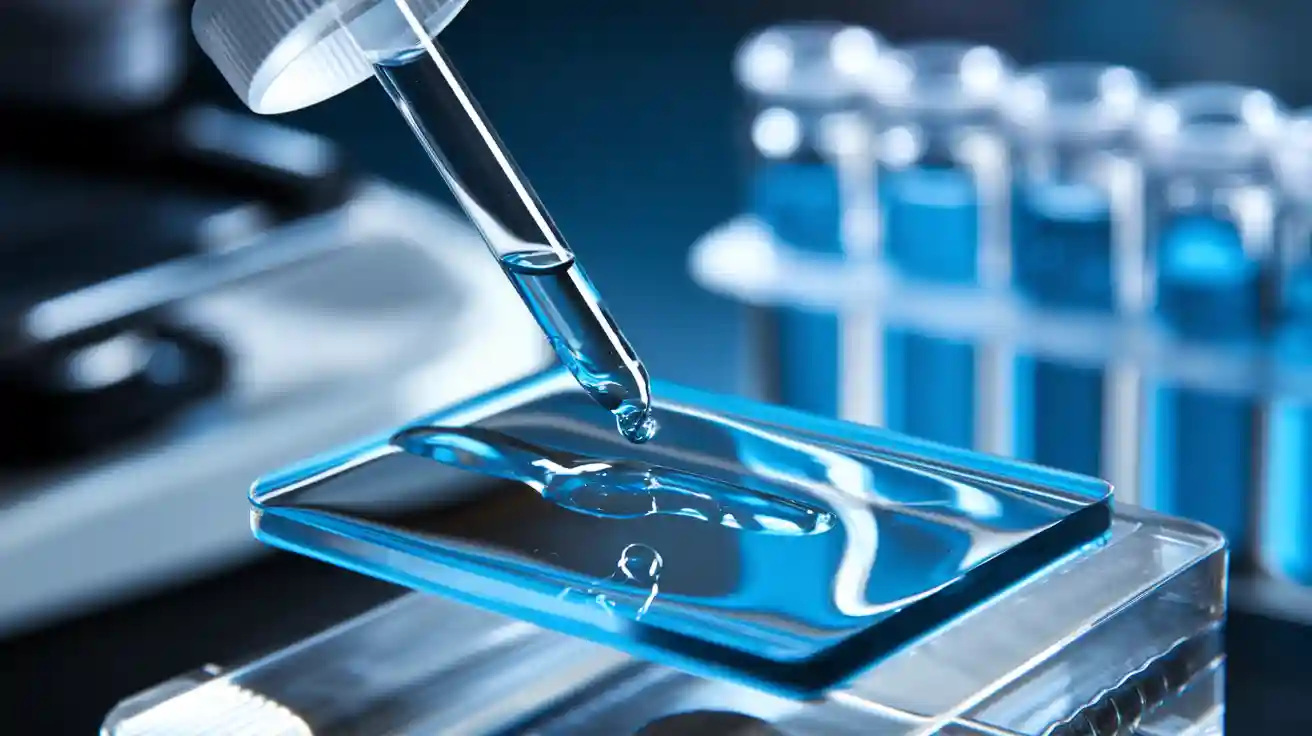
How to Stain a Microscope Slide
Preparing the Specimen for Staining
Placing the Specimen on the Slide
To begin staining a microscope slide, you must first prepare the specimen. Place the specimen carefully on the slide to ensure it is secure and ready for staining. If you are working with a thin tissue section, use a microtome to slice it evenly.
Using a pipette to transfer a small drop onto the slide for liquid samples. Spread the sample gently using a clean cover slip or applicator to create a thin, even layer. This step ensures the stain can penetrate the specimen effectively.
Ensuring Proper Placement for Staining
Proper placement of the specimen is critical for achieving clear results. Avoid overcrowding the slide, as this can lead to uneven staining. Ensure the specimen is centered and lies flat on the slide. If you are working with paraffin-embedded sections, make sure the slide is completely dewaxed. Residual wax can block the stain, resulting in patchy or inconsistent results.
Hydrate the specimen thoroughly before staining to prevent contamination and ensure the stain adheres evenly.
Applying the Stain Step by Step
Techniques for Even Application
Even application of the stain is essential for clear and consistent results. Use high-quality staining reagents to avoid variability. Maintain consistent staining conditions by controlling factors like temperature and humidity.
Use staining racks or jigs to ensure uniform application across all samples for multiple slides. These tools minimize human error and help you achieve professional-quality results.
|
Technique |
Description |
|---|---|
|
Use High-Quality Staining Reagents |
Invest in reliable staining kits and reagents to ensure consistent results. |
|
Maintain Consistent Staining Conditions |
Control factors like temperature, humidity, and staining time to achieve uniform staining. |
|
Staining Jigs and Racks |
Utilize staining jigs or racks to hold multiple slides, ensuring even staining and reducing the risk of human error. |
Timing and Adjustments for Best Results
Timing plays a crucial role in staining a microscope slide. Follow the staining protocol carefully and use a timer to ensure accuracy. Overstaining can obscure details, while understaining may fail to highlight important structures. After applying the stain, rinse the slide gently with distilled water to remove excess stain without damaging the specimen. Allow the slide to air-dry or use a drying tool before proceeding to the next step.
Note: Renew your staining reagents regularly to maintain their effectiveness. Old or contaminated reagents can lead to inconsistent results.
Rinsing and Drying Microscope Slides
Rinsing Off Excess Stain
How to Rinse Without Damaging the Specimen
Rinsing is a critical step in staining a microscope slide. It removes excess stain while preserving the integrity of your specimen. To avoid damage, always use gentle techniques.
High-purity water, such as deionized or distilled water, is ideal for rinsing. This prevents contaminants from interfering with your results. Hold the slide at an angle and let the water flow over it naturally.
Avoid using high-pressure streams, as they can dislodge or distort delicate specimens.
Tools and Techniques for Effective Rinsing
Using the right tools ensures effective rinsing without compromising your specimen. Here are some recommendations:
-
Deionized or distilled water for purity.
-
A gentle pipette or dropper for controlled water application.
-
A clean, lint-free cloth to absorb excess water if needed.
These tools help you maintain the quality of your specimen while removing unwanted stain residues.
Drying the Slide for Clear Results
Air Drying vs. Using a Drying Tool
Drying your slide properly is just as important as rinsing. Air drying in a clean, dust-free environment is a simple and effective method. However, it requires patience, as it can take time.
You can use a drying tool like a filtered air blower for faster results. This method minimizes the risk of introducing contaminants and ensures a completely dry surface.
Ensuring the Slide is Completely Dry
Follow these steps to ensure your slide is fully dry before use:
-
Rinse the slide thoroughly to remove any contaminants.
-
Place the slide in a clean, dust-free area for air drying, or gently pat it dry with a lint-free cloth.
-
Use a filtered air blower for critical applications to expedite drying and prevent contamination.
A completely dry slide ensures the stain remains intact and provides the clearest view of your specimen.
Tips for Achieving Clear Results with Stained Slides
Common Mistakes to Avoid
Even small errors can lead to poor results when staining a microscope slide. Avoiding these common mistakes will help you achieve clear and consistent outcomes:
-
Sanding Issues: Sanding the slide too much or too little can affect how well the stain adheres. Always ensure the surface is smooth but not overly polished.
-
Application Problems: Uneven application of stains can result in runs, pools, or laps. Use controlled techniques to apply the stain evenly across the slide.
-
Drying Time: Rushing the drying process can cause adhesion problems. Allow the slide to dry completely before proceeding to the next step.
-
Mixing Stain: Failing to mix the stain thoroughly can lead to inconsistent coloration. Stir or shake the stain solution well before use to ensure uniformity.
Paying attention to these details will save you time and effort while improving the quality of your microscopy results.
Best Practices for Consistent and Accurate Results
To achieve the best results when staining a microscope slide, follow these best practices:
-
Use high-quality microscopy stains and reagents. This ensures consistent and reliable results every time.
-
Maintain consistent staining conditions. Control factors like temperature, humidity, and staining time to avoid variability.
-
Utilize staining jigs or racks. These tools help you hold multiple slides securely, ensuring even application of stains.
-
Standardize your staining process. Implement standard operating procedures for reagent preparation, application, and timing.
-
Monitor quality regularly. Use control slides to check for consistency and accuracy throughout the process.
Adopting these practices allows you to produce slides with clear and detailed microscopy results. Consistency in your approach will also make it easier to replicate your findings in future experiments.
Staining microscope slides requires a systematic approach to achieve clear and accurate results. Follow these key steps to ensure success:
-
Preparation: Create wet mounts to position your specimen within the field of view.
-
Fixation: Use heat or chemical fixatives to preserve the shape and rigidity of cells or tissues.
-
Mordants: Apply chemical agents to help dyes bind to otherwise unstainable materials.
-
Permeabilization: Treat the specimen with surfactants to allow dye molecules to penetrate cell membranes.
Each step plays a vital role in enhancing the visibility of your specimen. Practicing these techniques will improve your skills and help you achieve consistent results. With time and attention to detail, you can master the art of slide staining and produce professional-quality slides every time.
FAQ
What is the purpose of staining microscope slides?
Staining enhances the contrast of your specimen, making structures more visible under the microscope. It allows you to identify specific components, such as cell walls or nuclei, that would otherwise remain transparent or difficult to observe.
How do you choose the right stain for a specimen?
Select a stain based on your specimen’s properties and the details you want to highlight. For example, use Gram staining for bacteria or eosin for cytoplasmic components. Always consider the compatibility of the stain with your fixation method.
Can you reuse staining solutions?
Reusing staining solutions is not recommended. Old or contaminated solutions can lead to inconsistent results. Always prepare fresh solutions to ensure the stain binds effectively and produces clear, accurate observations.
How do you avoid overstaining a slide?
Follow the staining protocol and use a timer to control the duration. Rinse the slide immediately after the recommended time to remove excess stain. Overstaining can obscure details, so monitoring the process is essential.
What should you do if the stain doesn’t adhere evenly?
Uneven staining often results from improper cleaning or placement of the specimen. Ensure the slide is clean and the specimen is flat and centered. Use high-quality stains and maintain consistent conditions during the staining process.

