
When I think about tools that bring the microscopic world to life, stereo microscopes stand out. These devices use two separate optical paths to deliver a three-dimensional view, making them indispensable for tasks requiring depth perception. Their dual-optical system allows each eye to see slightly different angles, creating a realistic 3D image. If you’re wondering how do stereo microscopes work, it’s this unique design that makes them perfect for examining larger, opaque specimens at low magnification.
They shine in fields like dissection, entomology, and botany, where observing fine details is crucial. Professionals also rely on them for tasks like inspecting circuit boards, analyzing fossils, or identifying minerals. Whether in forensics or manufacturing, stereo microscopes provide clarity and precision.
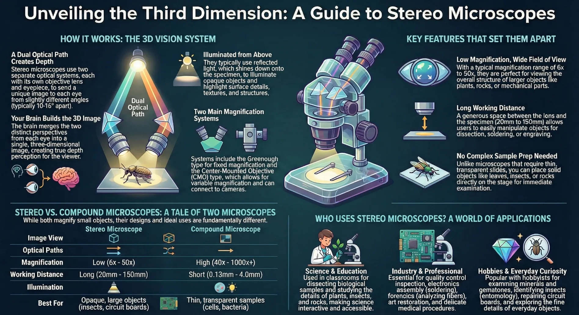
How Do Stereo Microscopes Work?
The Dual Optical Path System
When I first learned about stereo microscopes, their dual optical path system fascinated me. This system uses two separate optical paths, each with its own objective lens and eyepiece. This design allows each eye to view the specimen from slightly different angles, creating a three-dimensional image. The Greenough principle, a key concept in stereo microscope design, positions two identical optical systems at an angle of 10° to 16°. This setup enhances depth perception and makes it easier to observe fine details.
Modern stereo microscopes often feature a shared main objective with parallel beam paths. This design, introduced in 1957 by the American Optical Company, includes a five-step magnification changer. It’s incredible how these advancements have improved the way we view specimens.
How 3D Imaging is Achieved
I’ve always found the three-dimensional imaging capability of stereo microscopes remarkable. This effect is achieved by directing two optical paths to create distinct viewing angles for each eye. The result is a realistic 3D visualization of the specimen. There are two main types of magnification systems in stereo microscopes: the Greenough type, which offers fixed magnification, and the center-mounted objective (CMO) type, which allows for variable magnification.
The CMO type is versatile. It can connect to CCD cameras, enabling the capture of stereoscopic video images. Beam splitters in the optical paths further enhance this capability, making it possible to record and analyze 3D images.
The Role of Light and Magnification
Light and magnification play crucial roles in how a stereo microscope works. Unlike compound microscopes, which have higher magnification ranges (40x to 100x), stereo microscopes operate at lower magnifications, typically between 6x and 50x. This range is perfect for viewing larger specimens or examining details without extreme zoom.
Stereo microscopes also use reflected light to illuminate opaque objects. This lighting method highlights surface details, making it easier to study textures and structures.
The combination of lower magnification and reflected light ensures that the three-dimensional image remains clear and detailed.
Unique Features of Stereo Microscopes
3D Imaging and Depth Perception
One of the most fascinating features of stereo microscopes is their ability to provide a three-dimensional view. Unlike conventional microscopes, which display flat, two-dimensional images, stereo microscopes use two separate optical paths. Each path has its own objective lens and eyepiece, creating slightly different perspectives for each eye. This design allows the brain to merge these perspectives into a single 3D image.
I’ve noticed that this depth perception is especially useful when examining solid objects like fossils, insects, or circuit boards. Technologies like FusionOptics take this even further. One optical path offers high resolution, while the other provides a greater depth of field. Together, they create a detailed and immersive view of the specimen.
Adjustable magnification and working distance enhance this experience, making it easier to focus on intricate details.
Low Magnification for Larger Specimens
Stereo microscopes excel at observing larger specimens. Their low magnification range, typically between 6x and 50x, provides a broader field of view. This makes it easier to study the overall structure of objects like plants, rocks, or mechanical components.
High magnification, while useful for smaller details, often reduces clarity and limits the visible area.
I’ve found that this low magnification is perfect for tasks requiring a comprehensive view. For example, when analyzing a gemstone, I can see its entire surface without losing focus on its finer details. This balance between clarity and field of view is what sets stereo microscopes apart from other optical instruments.
Reflected Light for Opaque Objects
Another unique feature of stereo microscopes is their use of reflected light. Unlike transmitted light, which passes through transparent specimens, reflected light illuminates opaque objects from above. This lighting method enhances surface details and maximizes contrast, making textures and structures more visible. I’ve used this feature extensively when examining materials like metals, fabrics, or ceramics. The ability to see surface imperfections or patterns clearly is invaluable in fields like manufacturing and quality control.
Reflected light ensures that even the smallest details stand out, providing a level of precision that other microscopes can’t match.
Stereo Microscopes vs. Compound Microscopes
Differences in Optical Design
When comparing stereo microscopes and compound microscopes, their optical designs stand out as fundamentally different. Stereo microscopes use two separate optical paths, creating a three-dimensional view. This design provides depth perception, making them ideal for tasks requiring spatial awareness. In contrast, compound microscopes rely on a single optical path, producing a flat, two-dimensional image.
Magnification capabilities also differ significantly. Compound microscopes offer higher magnification, ranging from 40x to 1000x. This makes them suitable for observing microscopic details like cells or bacteria. Stereo microscopes, on the other hand, operate at lower magnifications, typically between 6x and 50x. This range is perfect for examining larger specimens.
Working distance is another key difference. Stereo microscopes have longer working distances, often between 20mm and 150mm. This allows me to manipulate specimens like circuit boards or fossils with ease.
Compound microscopes, with their shorter working distances (0.13mm to 4.0mm), are better suited for thin, transparent samples.
Applications of Each Type
I’ve noticed that stereo microscopes excel in fields where depth perception and surface detail are crucial. They’re indispensable for tasks like dissection, gemology, and forensics. I’ve also used them for examining minerals and performing intricate tasks like engraving or dental work. Compound microscopes, however, shine in biological studies. They’re perfect for observing cells, tissues, and microorganisms. Their high magnification and fine focus adjustments make them essential in medical research and microbiology.
Advantages and Limitations
Each type of microscope has its strengths and weaknesses. Stereo microscopes offer a three-dimensional view and longer working distances, making them versatile for larger specimens. However, their lower magnification limits their ability to observe very small details. Even with auxiliary lenses, they can’t match the 1000x magnification of compound microscopes. Compound microscopes provide unparalleled detail at high magnifications. This makes them ideal for studying microscopic structures.
However, their two-dimensional imaging and shorter working distances make them less practical for larger, opaque specimens.
Applications of Stereo Microscopes
Scientific and Educational Uses
I’ve seen stereo microscopes transform the way students and researchers explore the natural world. In classrooms, digital stereo microscopes make learning interactive. They allow students to observe samples in detail while projecting images onto screens for group discussions. This technology simplifies teaching and encourages collaboration. What I find most remarkable is how user-friendly these microscopes are for young learners.
Unlike other types, stereo microscopes don’t require complex sample preparation. Students can directly examine natural specimens like leaves, insects, or rocks. This hands-on approach fosters curiosity and makes science more accessible.
Industrial and Professional Applications
Stereo microscopes are essential tools in many industries. I’ve used them in quality control to inspect small parts for defects. Their 3D imaging and depth perception make them perfect for tasks requiring precision. In electronics, they’re invaluable for soldering and circuit board inspection. Industries like automotive and aerospace rely on stereo microscopes for assembly and inspection processes.
These microscopes also play a significant role in healthcare. Surgeons and diagnosticians use them to examine tissues and perform delicate procedures. Their versatility extends to museums and art galleries, where they help preserve and study artifacts.
Here’s a quick look at some common applications:
-
Dissecting plant and animal samples in academic settings.
-
Observing surface details in botany, geology, and gemology.
-
Directly interacting with materials in industrial tasks.
Everyday and Hobbyist Uses
Stereo microscopes aren’t just for professionals. I’ve seen hobbyists use them to explore their passions. For example, they’re ideal for examining minerals, fossils, and gemstones. Enthusiasts in electronics often use them to repair circuit boards or inspect components. In my experience, these microscopes are also popular among entomologists. It’s easy to study insects in detail with these tools. From dissecting tiny organisms to analyzing complex patterns, a stereo microscope unlocks a world of possibilities.
For everyday use, they’re perfect for anyone curious about the small details of the world around them. Their simplicity and versatility make them a great addition to any hobbyist’s toolkit. I’ve seen their versatility firsthand across various fields. In biology, they simplify dissections and the study of model organisms. In geology, they reveal intricate details of minerals and fossils. Forensics relies on them for analyzing fibers and hair, while manufacturing uses them for quality control. Their 3D imaging capability also supports art restoration and environmental studies.
Understanding how stereo microscopes work highlights their importance. The dissecting microscope, with its dual optical paths, offers a unique perspective that enhances observation. Its use of reflected light and low magnification ensures detailed examination of larger specimens. This combination of features makes the dissecting microscope a vital tool in science, industry, and even hobbies.
From classrooms to laboratories, the dissecting microscope continues to inspire curiosity and innovation. Its role in education, research, and professional applications underscores its value. I believe this tool will remain essential for anyone exploring the fine details of the world around us.

