
Have you ever wondered what electron microscopes are used for? These powerful tools have revolutionized scientific research, offering a window into the microscopic world. While many are familiar with their roles in traditional fields like biology and materials science, you might be surprised by some lesser-known applications.
From diagnostic applications to the development of new materials, electron microscopy opens up a world of possibilities. This blog will help you uncover the remarkable applications of electron microscopy that you may not yet know.
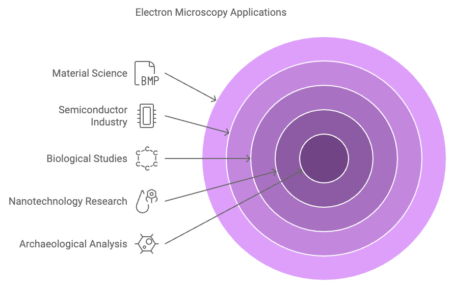
Use Case 1: Nanotechnology Research
Have you ever marveled at the tiny wonders of the world? Imagine peering into a realm where materials are so small that they defy imagination. Welcome to the world of nanotechnology, where electron microscopes play a starring role.
These powerful tools have become indispensable in advancing technology and providing unique insights into nanoscale materials.
Importance in Advancing Technology
Nanotechnology is not just a buzzword; it’s a revolution. Electron microscopes have become the backbone of this field, enabling scientists to explore and manipulate materials at the atomic level. This capability is crucial for developing new technologies that can transform industries. From enhancing energy efficiency to solving health problems, the applications of nanotechnology are vast and varied.
Unique Insights into Nanoscale Materials
Electron microscopes offer a window into the nanoscale world, revealing invisible details to the naked eye. They allow researchers to study nanoparticles with unprecedented clarity, leading to breakthroughs in various fields.
For instance, scientists have discovered that nanoparticles can exhibit distinct colors, paving the way for multicolor electron microscopy in the future. This insight is just the tip of the iceberg in understanding the complex behavior of nanoscale materials.
What You Need to Know?
Basic Principles of Nanotechnology
Before we take a closer look at electron microscopy, it’s essential to grasp the basics of nanotechnology. This field involves manipulating materials at the nanoscale, typically between 1 and 100 nanometers. At this scale, materials can exhibit unique properties that differ from their bulk counterparts.
Understanding these principles is key to unlocking the potential of electron microscopes in nanotech research.
Role of Electron Microscopes in Nanotech
Electron microscopes are the workhorses of nanotechnology research. They provide the high-resolution images needed to study the structure and composition of nanoscale materials. By using electron beams instead of light, these microscopes achieve far greater resolutions, allowing scientists to observe the intricate details of nanoparticles.
This capability is vital for developing new materials and understanding their properties.
Step by Step Instructions
Preparing Samples
Sample preparation is a critical step in electron microscopy. You’ll need to ensure that your samples are thin enough for the electron beam to pass through. This often involves cutting or slicing the material to the desired thickness.
Proper preparation is essential for obtaining clear and accurate images.
Operating the Microscope
Once your samples are ready, it’s time to operate the electron microscope. Begin by placing the sample in the microscope’s chamber. Adjust the settings to focus the electron beam on the area of interest.
As you capture images, pay attention to the details and take notes on any observations. With practice, you’ll become adept at navigating the nanoscale world.
Potential Challenges
Have you ever tried to bake a cake and ended up with a lopsided mess? Well, preparing samples for an electron microscope can feel just as tricky! Sample preparation is a meticulous process. You must ensure that your samples are thin enough for the electron beam to pass through without distortion.
This often involves slicing the material to a precise thickness, which can be challenging, especially with delicate or complex materials. Once you’ve got your sample ready, interpreting the results is the next hurdle. The images produced by an electron microscope are incredibly detailed, but they require a trained eye to decipher. Misinterpretation can lead to incorrect conclusions, much like mistaking salt for sugar in your cake recipe!
Pro Tips
To navigate these challenges, here are some pro tips that can make your journey smoother:
-
Best Practices for Sample Handling: Always handle your samples with care. Use clean tools and work in a dust-free environment to avoid contamination. Think of it as handling a fragile piece of art.
-
Tips for Accurate Imaging: Calibration is key. Regularly calibrate your electron microscope to ensure accuracy. Adjust the focus and magnification settings carefully to capture the best possible image. It’s like tuning a guitar before a performance – precision matters!
Common Mistakes to Avoid | Pitfalls
Even seasoned researchers can stumble upon common pitfalls. Here are a couple of mistakes to watch out for:
-
Overlooking Calibration: Skipping calibration is like driving a car with misaligned wheels. It can lead to skewed results and wasted time. Make calibration a routine part of your process.
-
Misinterpreting Data: The images from an electron microscope can be complex. Avoid jumping to conclusions without thorough analysis. Take your time to study the details and consult with peers if needed. Remember, patience is a virtue!
Bottomline – Is This Use Case for You?
If you’re a researcher in this field, you’ll find these tools indispensable. They offer unparalleled insights into nanoscale materials, helping you push the boundaries of innovation. When you’re developing new materials or exploring energy solutions, electron microscopes can be your best allies.
Suitability for Researchers in Nanotechnology
For seasoned researchers, electron microscopes provide the precision needed to explore nanoparticles. These instruments have become essential in understanding chemical and structural characteristics at the nanoscale. Imagine the possibilities: enhancing energy efficiency, solving health problems, and even cleaning the environment. The electron microscope is your gateway to these advancements.
Considerations for Beginners
If you’re starting, don’t worry! Electron microscopes might seem complex, but they’re accessible with practice. Begin by familiarizing yourself with the basics of nanotechnology. Understand how these microscopes work and their role in your research. You’ll soon find that they open up a world of possibilities, allowing you to explore the microscopic universe with confidence.
Use Case 2: Biological Studies
Have you ever peered through a microscope and marveled at the intricate world of cells? Imagine diving even deeper, exploring the tiniest details of cellular structures with an electron microscope. This powerful tool is a game-changer in biological studies, offering insights that are crucial for medical research and beyond.
Why I Recommend It?
Enhances Understanding of Cellular Structures
You might wonder how scientists unravel the mysteries of life at the cellular level. The electron microscope is your answer. It allows you to see cellular structures in stunning detail, revealing the complex architecture of cells. This understanding is vital for studying diseases, developing treatments, and advancing our knowledge of biology.
Crucial for Medical Research
When it comes to medical research, precision is everything. Electron microscopes provide the clarity needed to examine tissues, cells, and organelles. They play a pivotal role in understanding diseases at the molecular level, aiding in the development of new therapies.
Imagine the impact of discovering a new treatment for a disease by observing its cellular behavior!
What You Need to Know?
Basics of Cell Biology
Before we go further, let’s grasp the basics of cell biology. Cells are the building blocks of life, each with unique functions and structures. Understanding these fundamentals helps you appreciate the detailed images you’ll capture with an electron microscope.
Application of Electron Microscopy in Biology
Electron microscopy is a cornerstone in biological research. It allows you to visualize the ultrastructure of cells, providing insights into their function and behavior. This application is essential for studying everything from viruses to complex tissues, offering a window into the microscopic world.
Step by Step Instructions
Sample Preparation for Biological Specimens
Preparing biological samples requires precision. You’ll need to fix and dehydrate your specimens to preserve their structure. This process often involves using chemicals to stabilize the cells, ensuring they remain intact during imaging. Proper preparation is key to obtaining clear and accurate images.
Imaging Techniques
Once your samples are ready, it’s time to capture images. Place the specimen in the electron microscope and adjust the settings to focus on the area of interest. Use different imaging techniques to highlight specific structures, such as staining to enhance contrast.
With practice, you’ll master the art of capturing stunning images that reveal the secrets of life.
Potential Challenges
Have you ever tried to preserve a delicate flower, only to find it wilted and lifeless the next day? Preserving biological samples for electron microscopy can feel just as challenging. You must maintain the integrity of your specimens to achieve high-resolution images. This process involves meticulous preparation and handling.
Preserving Biological Samples
Preserving biological samples requires precision and care. You need to fix and dehydrate your specimens to maintain their structure. This often involves using chemicals like formaldehyde or glutaraldehyde to stabilize the cells. Think of it as embalming a specimen to keep it in pristine condition for examination under the electron microscope.
Without proper preservation, your samples may degrade, leading to inaccurate results.
Achieving High-Resolution Images
Achieving high-resolution images with an electron microscope is no small feat. You must ensure that your samples are thin enough for the electron beam to penetrate without distortion. This often involves slicing the material to a precise thickness.
The challenge lies in balancing the need for thin samples with the risk of damaging delicate structures. It’s like trying to slice a cake into paper-thin layers without crumbling it.
Pro Tips
Navigating these challenges can be daunting, but with the right techniques, you can master the art of electron microscopy.
Techniques for Sample Preservation
-
Use Appropriate Fixatives: Choose the right fixative for your sample type. For instance, formaldehyde works well for preserving proteins, while glutaraldehyde is better for lipids.
-
Control Temperature and Humidity: Maintain a stable environment to prevent sample degradation. Think of it as creating a climate-controlled room for your specimens.
Tips for Optimizing Image Quality
-
Calibrate Regularly: Regular calibration ensures your electron microscope produces accurate images. It’s like tuning a musical instrument before a performance.
-
Adjust Beam Settings: Experiment with different beam settings to find the optimal balance between resolution and contrast. This approach helps you capture the finest details without losing clarity.
Common Mistakes to Avoid | Pitfalls
Even seasoned researchers can fall into common pitfalls when working with electron microscopes. Here are some mistakes to watch out for:
Inadequate Sample Fixation
Inadequate fixation can lead to sample degradation and poor image quality. Ensure you use the correct fixative and follow the recommended protocols. It’s like building a house on a shaky foundation—without proper fixation, your results may crumble.
Ignoring Environmental Conditions
Ignoring environmental conditions can compromise your samples. Maintain a stable temperature and humidity level to prevent artifacts from forming. Think of it as protecting a delicate painting from the elements.
Bottomline – Is This Use Case for You?
Have you ever imagined peering into the intricate world of cells and tissues with an electron microscope? If you’re a biologist or medical researcher, this tool is your gateway to groundbreaking discoveries. The electron microscope offers unparalleled insights into cellular structures, making it indispensable for those in the field. You can explore the molecular nature of diseases, develop new treatments, and even uncover the secrets of life itself.
Ideal for Biologists and Medical Researchers
For biologists and medical researchers, the electron microscope is a game-changer. It allows you to visualize cells and tissues in stunning detail, providing a deeper understanding of biological processes. Whether you’re studying viruses, bacteria, or complex tissues, the electron microscope reveals the hidden intricacies that are crucial for your research.
Discovering new treatments by studying cell behavior at the molecular level can revolutionize medical science. This tool empowers you to advance healthcare significantly.
Considerations for Non-Specialists
If you’re not a specialist, don’t worry! The electron microscope might seem daunting, but it’s accessible with practice. Start by familiarizing yourself with the basics of cell biology and electron microscopy. Understand how these microscopes work and their role in research.
Use Case 3: Material Science
Have you ever wondered how the materials around you are designed and perfected? Imagine having the power to see the tiniest details of a material’s structure. That’s where the electron microscope comes into play, revolutionizing material science.
Why I Recommend It?
Essential for Understanding Material Properties
You need to understand the properties of materials to innovate and improve them. Electron microscopes allow you to explore materials at an atomic level, revealing their secrets. This insight is crucial for industries like aerospace and electronics, where precision matters.
Supports Innovation in Material Design
Innovation thrives on understanding. By using an electron microscope, you can identify faults in materials before they become problems. This capability supports the creation of stronger, more efficient materials, paving the way for groundbreaking advancements.
What You Need to Know?
Fundamentals of Material Science
Material science is all about studying the properties and applications of materials. You’ll explore how materials behave under different conditions and how they can be manipulated to serve specific purposes. This knowledge forms the backbone of using electron microscopy effectively.
Role of Electron Microscopy in Material Analysis
Electron microscopy provides high-resolution images that are essential for analyzing materials. By visualizing materials at such a fine scale, you can understand their composition and structure.
This understanding helps in quality control and failure analysis, ensuring safety and reliability.
Step by Step Instructions
Preparing Material Samples
-
Select the Material: Choose the material you want to analyze. Consider its properties and the information you seek.
-
Cut to Size: Slice the material into thin sections. This step ensures the electron beam can penetrate and provide clear images.
-
Clean the Sample: Remove any contaminants. Clean samples lead to more accurate results.
Conducting Analysis
-
Place in Microscope: Insert the prepared sample into the electron microscope.
-
Adjust Settings: Focus the electron beam on the area of interest. Adjust magnification to capture detailed images.
-
Analyze Results: Study the images to understand the material’s structure. Look for patterns or anomalies that could affect performance.
Potential Challenges
Have you ever tried juggling different tasks at once? Handling diverse materials with an electron microscope can feel just as tricky. Each material has unique properties, requiring specific preparation techniques. You must adapt your approach to ensure accurate results. For instance, metals might need polishing, while biological samples require careful dehydration. This diversity demands flexibility and expertise.
Ensuring accurate measurements is another hurdle. The electron microscope offers incredible detail, but precision is key. Misjudging measurements can lead to incorrect conclusions. You need to calibrate the microscope regularly and pay close attention to settings. Think of it like tuning a musical instrument; even a slight error can disrupt harmony.
Pro Tips
To navigate these challenges, consider these strategies:
-
Strategies for Sample Preparation: Tailor your preparation method to the material. Metals may need polishing, while organic samples might require special coatings. Always clean your samples thoroughly to avoid contamination.
-
Tips for Precise Analysis: Regularly calibrate your electron microscope. Adjust the focus and magnification carefully. Use reference standards to verify accuracy. This approach ensures that your analysis remains reliable and consistent.
Common Mistakes to Avoid | Pitfalls
Even seasoned users can stumble upon common pitfalls. Here are some mistakes to watch out for:
-
Overlooking Material Properties: Ignoring the unique characteristics of materials can lead to errors. Always consider the specific needs of each sample. This awareness helps you choose the right preparation and imaging techniques.
-
Misjudging Measurement Accuracy: Failing to calibrate or adjust settings can skew results. Double-check your measurements and consult with peers if needed. Precision is crucial for meaningful insights.
Bottomline – Is This Use Case for You?
Have you ever wondered if you could unlock the secrets of materials with an electron microscope? If you’re a material scientist or engineer, this tool is your gateway to innovation. Electron microscopes allow you to explore materials at the atomic level, revealing insights that drive advancements in industries like aerospace and electronics.
Suitable for Material Scientists and Engineers
You, as a material scientist, will find electron microscopes indispensable. They provide the precision needed to understand material properties and support innovation in design. Imagine identifying faults before they become problems, ensuring safety and reliability in your projects. The electron microscope is your ally in achieving these goals.
Key Benefits for Material Scientists:
-
High Resolution: Visualize materials with incredible detail.
-
Quality Control: Identify potential faults early.
-
Innovation Support: Develop new materials with enhanced properties.
The electron microscope is more than just a tool; it’s a window into the microscopic world. It does not matter if you’re a seasoned researcher or a curious beginner, this powerful instrument offers endless opportunities for exploration and discovery.
Use Case 4: Semiconductor Industry
Have you ever wondered how your smartphone gets smarter every year? The secret lies in the semiconductor industry, where electron microscopes play a pivotal role. These powerful tools are essential for quality control and innovation, ensuring that the tiny components inside your devices work flawlessly.
Why I Recommend It?
Critical for Quality Control and Innovation
In the semiconductor world, precision is everything. You need to ensure that every chip functions perfectly. Electron microscopes allow you to inspect these tiny structures with incredible detail. They help identify defects that could lead to failures, saving time and resources.
This capability supports continuous innovation, pushing the boundaries of what’s possible in electronics.
Supports Advancements in Electronics
The rapid evolution of technology relies on advancements in semiconductors. Electron microscopes provide the insights needed to develop smaller, faster, and more efficient components.
By examining materials at the atomic level, you can discover new ways to enhance performance and reduce energy consumption.
What You Need to Know?
Basics of Semiconductor Technology
Semiconductors are the building blocks of modern electronics. They control the flow of electricity in devices, enabling everything from computers to smartphones. Understanding their basic properties helps you appreciate the role of electron microscopy in this field.
Application of Electron Microscopy in Semiconductors
Electron microscopes are indispensable in semiconductor research. They offer high-resolution images that reveal the intricate details of semiconductor materials. This information is crucial for designing and manufacturing cutting-edge components.
Step by Step Instructions
Sample Preparation for Semiconductor Materials
-
Select the Material: Choose the semiconductor material you want to analyze.
-
Cut to Size: Slice the material into thin sections to allow electron penetration.
-
Clean the Sample: Remove any contaminants to ensure accurate imaging.
Imaging and Analysis Techniques
-
Place in Microscope: Insert the prepared sample into the electron microscope.
-
Adjust Settings: Focus the electron beam on the area of interest. Adjust magnification for detailed images.
-
Analyze Results: Study the images to understand the material’s structure. Look for defects or patterns that could impact performance.
Potential Challenges
Have you ever tried assembling a tiny model ship inside a bottle? Handling delicate semiconductor structures with an electron microscope can feel just as intricate. You must navigate these tiny landscapes with precision. Each component is fragile, and even the slightest misstep can lead to damage.
Handling Delicate Semiconductor Structures
Semiconductors are like the fine china of the tech world. You need to handle them with care. The electron microscope allows you to see these structures in incredible detail, but it requires a gentle touch. Avoid applying too much pressure or using improper tools.
Think of it as performing surgery on a microchip.
Ensuring Precision in Analysis
Precision is key when analyzing semiconductor materials. The electron microscope provides high-resolution images, but interpreting them accurately is crucial. Misinterpretation can lead to costly errors.
Regular calibration and careful attention to settings ensure that your analysis remains spot-on.
Pro Tips
Navigating the world of semiconductors with an electron microscope can be challenging, but these pro tips will help you maintain precision and clarity.
Best Practices for Semiconductor Imaging
-
Use Clean Samples: Always start with clean, contaminant-free samples. This practice ensures clear imaging and accurate results.
-
Regular Calibration: Keep your electron microscope calibrated. Regular checks prevent errors and maintain image quality.
Tips for Maintaining Precision
-
Adjust Beam Settings: Fine-tune the electron beam settings to match the material’s needs. This adjustment helps capture the finest details without distortion.
-
Consult with Peers: Collaborate with colleagues to verify findings. A second opinion can provide new insights and prevent mistakes.
Common Mistakes to Avoid | Pitfalls
Even experienced users can fall into common traps. Here are some pitfalls to watch out for:
Neglecting Sample Integrity
Neglecting sample integrity is like trying to paint on a wet canvas. Ensure your samples are well-prepared and stable. This preparation prevents artifacts and ensures accurate imaging.
Misinterpreting Semiconductor Data
Misinterpreting data can lead to incorrect conclusions. Take your time to analyze the images thoroughly. Consult with peers if needed. Remember, patience and collaboration are your best allies.
Bottomline – Is This Use Case for You?
Have you ever imagined peering into the tiny world of semiconductors with an electron microscope? If you’re in the semiconductor industry, this tool is your best friend. It offers precision and clarity, essential for ensuring quality and driving innovation.
Ideal for Professionals in the Semiconductor Industry
You, as a professional, will find the electron microscope indispensable. It allows you to inspect semiconductor materials with unmatched detail. This capability helps you identify defects early, saving time and resources. Imagine the satisfaction of knowing that every chip functions perfectly because of your meticulous analysis.
Benefits for Professionals:
-
High Precision: Capture intricate details of semiconductor structures.
-
Quality Assurance: Detect defects before they become costly issues.
-
Innovation Support: Develop cutting-edge components with enhanced performance.
Considerations for Newcomers
If you’re new to the field, don’t worry! The electron microscope might seem complex, but with practice, you’ll master it. Start by understanding the basics of semiconductor technology. Learn how these microscopes work and their role in your research. You’ll soon find that they open up a world of possibilities, allowing you to explore the microscopic universe with confidence.
Tips for Newcomers:
-
Learn the Basics: Familiarize yourself with semiconductor fundamentals.
-
Practice Regularly: Gain hands-on experience with the microscope.
-
Seek Guidance: Collaborate with experienced colleagues for insights.
Use Case 5: Archaeological Analysis
Have you ever held an ancient artifact and wondered about its story? Imagine having the power to uncover secrets hidden for centuries. With an electron microscope, you can explore the intricate details of historical materials, offering once unimaginable insights.
Why I Recommend It?
Offers Insights into Ancient Artifacts
You can delve into the past with an electron microscope, revealing the craftsmanship and materials used by ancient civilizations. This tool allows you to examine artifacts at a microscopic level, uncovering details invisible to the naked eye.
For instance, you might discover the specific minerals used in a pottery glaze or the wear patterns on a tool, providing clues about its use and origin.
Enhances Understanding of Historical Materials
Understanding the composition of historical materials is crucial for preservation and restoration. An electron microscope helps you identify the elements and compounds present in artifacts, guiding conservation efforts. This knowledge ensures that these treasures are preserved for future generations, maintaining their historical integrity.
What You Need to Know?
Basics of Archaeology
Archaeology is the study of human history through artifacts and structures. You explore past cultures by analyzing the materials they left behind. This field requires a keen eye for detail and a passion for discovery.
Understanding the basics of archaeology helps you appreciate the significance of each artifact you examine.
Role of Electron Microscopy in Artifact Analysis
Electron microscopy plays a vital role in artifact analysis. It provides high-resolution images that reveal the microstructure of materials. This capability allows you to identify the techniques and materials used by ancient artisans.
Knowing these details helps you build the artifact’s story, revealing the lives of its creators.
Step by Step Instructions
Preparing Archaeological Samples
-
Select the Artifact: Choose an artifact that requires analysis. Consider its material and historical significance.
-
Clean the Sample: Gently clean the artifact to remove dirt and debris. Use soft brushes and mild solutions to avoid damage.
-
Cut to Size: If necessary, carefully cut a small section of the artifact for analysis. Ensure the sample is thin enough for electron penetration.
Conducting Detailed Analysis
-
Place in Microscope: Insert the prepared sample into the electron microscope.
-
Adjust Settings: Focus the electron beam on the area of interest. Adjust magnification to capture detailed images.
-
Analyze Results: Study the images to understand the artifact’s composition. Look for patterns or anomalies that could provide historical insights.
Potential Challenges
Have you ever tried to handle a delicate piece of ancient pottery, only to feel the anxiety of possibly breaking it? Preserving fragile artifacts with an electron microscope can feel just as nerve-wracking. You must ensure that these treasures remain intact during analysis.
The process requires a gentle touch and meticulous care. Even the slightest mishandling can lead to irreversible damage.
Preserving Fragile Artifacts
-
Handle with Care: Always use soft tools and gloves. This prevents oils and dirt from contaminating the artifact.
-
Stabilize the Environment: Maintain consistent temperature and humidity. Fluctuations can cause materials to expand or contract, leading to cracks.
-
Use Proper Supports: Secure the artifact with appropriate supports to prevent movement during analysis.
Interpreting Historical Data
Interpreting historical data can be like piecing together a complex puzzle. The electron microscope provides detailed images, but understanding them requires a keen eye. You must consider the context and historical background of each artifact.
Misinterpretation can lead to incorrect conclusions about its origin or use.
Pro Tips
Navigating the world of archaeological analysis with an electron microscope can be challenging, but these pro tips will help you preserve artifacts and interpret data accurately.
Techniques for Artifact Preservation
-
Use Gentle Cleaning Methods: Opt for soft brushes and mild solutions. This approach minimizes the risk of damaging delicate surfaces.
-
Implement Controlled Storage: Store artifacts in climate-controlled environments. This practice prevents deterioration over time.
Tips for Accurate Historical Analysis
-
Cross-Reference Findings: Compare your results with existing research. This helps validate your interpretations and provides a broader context.
-
Consult Experts: Collaborate with historians or archaeologists. Their insights can offer valuable perspectives on your findings.
Common Mistakes to Avoid | Pitfalls
Even seasoned researchers can stumble upon common pitfalls. Here are some mistakes to watch out for:
Damaging Artifacts During Preparation
Avoid using harsh chemicals or abrasive tools. These can cause irreversible damage. Always opt for the gentlest methods available.
Misreading Historical Context
Jumping to conclusions without thorough analysis can lead to errors. Take your time to study the details and consult with experts if needed. Remember, patience is key!
Bottomline – Is This Use Case for You?
Have you ever held an ancient artifact and felt the weight of history in your hands? Imagine using an electron microscope to uncover secrets hidden for centuries. This tool offers archaeologists and historians a unique opportunity to explore the past with precision and clarity.
Suitable for Archaeologists and Historians
You, as an archaeologist or historian, will find the electron microscope indispensable. It allows you to examine artifacts at a microscopic level, revealing details invisible to the naked eye. This capability helps you understand the materials and techniques used by ancient civilizations. Imagine discovering the specific minerals in a pottery glaze or the wear patterns on a tool. These insights can provide clues about an artifact’s use and origin.
Benefits for Archaeologists and Historians:
-
Detailed Analysis: Examine artifacts with incredible detail.
-
Material Identification: Identify elements and compounds in historical materials.
-
Preservation Guidance: Inform conservation efforts with precise data.
Considerations for Interdisciplinary Studies
If you’re venturing into archaeology from another field, don’t worry! The electron microscope might seem complex, but it’s accessible with practice. Start by understanding the basics of archaeology and how electron microscopy can enhance your research. You’ll soon find that these tools open up a world of possibilities, allowing you to explore the microscopic universe with confidence.
Tips for Interdisciplinary Researchers:
-
Learn the Basics: Familiarize yourself with archaeological fundamentals.
-
Practice Regularly: Gain hands-on experience with the microscope.
-
Collaborate: Work with experienced colleagues to gain insights.
The electron microscope is more than just a tool; it’s a window into the past. For both the adept researcher and the curious newcomer, this robust instrument affords infinite avenues for exploration and discovery.

